Not quite all back to in-person – the EFEM student Symposium 2022

Despite our last blog post celebrating the regained opportunity to meet with other researchers in person and all the benefits that come with it I just had the pleasure of presenting at the first European Federation for Experimental Morphology (EFEM) Student Symposium online.

While it would have been easy for me to attend in person, as the event was hosted and organised here at RCSI, not many others would have had it quite so easy. As the name suggests, researchers from all across Europe attended. Every EFEM associated anatomical society across Europe and RCSI, as the host institution, had the opportunity to select two members to present. I was honoured to be chosen to represent RCSI. Overall, 15 different countries were represented in the student talks which made for a diverse mix that was particularly nice for the bit of organized fun at the end of the first day which encouraged networking.

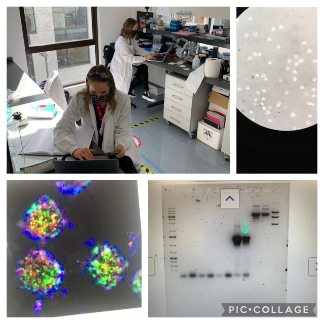
Especially, because having only begun my PhD this past year I felt the category Preliminary Results and Outlook aimed at Undergrads, Masters and early-stage PhD students perfectly suited the stage of my project. This was also a brilliant way to see what other students at my level were doing in the field of anatomical research all across Europe. Having this chance to see research in progress was refreshing and uplifting contrasted with the usually more rounded later stage presentations. Having studied anatomy in my undergraduate degree I was also delighted to simply engage with more conventional anatomical research than I currently do myself.

Ronja Struck, 1Yr PhD student at the EFEM Student Symposium 2022

A wonderful opportunity to gain insights into, for example, the implicit knowledge of academia was the career development part of the conference. Talks about academia, industry and publishing offered a chance to get an inside view of those career paths at different positions within them. Especially the typical day in a research journal’s editor provided a new perspective on what is important when writing papers and will have lasting benefits for me and my scientific writing.

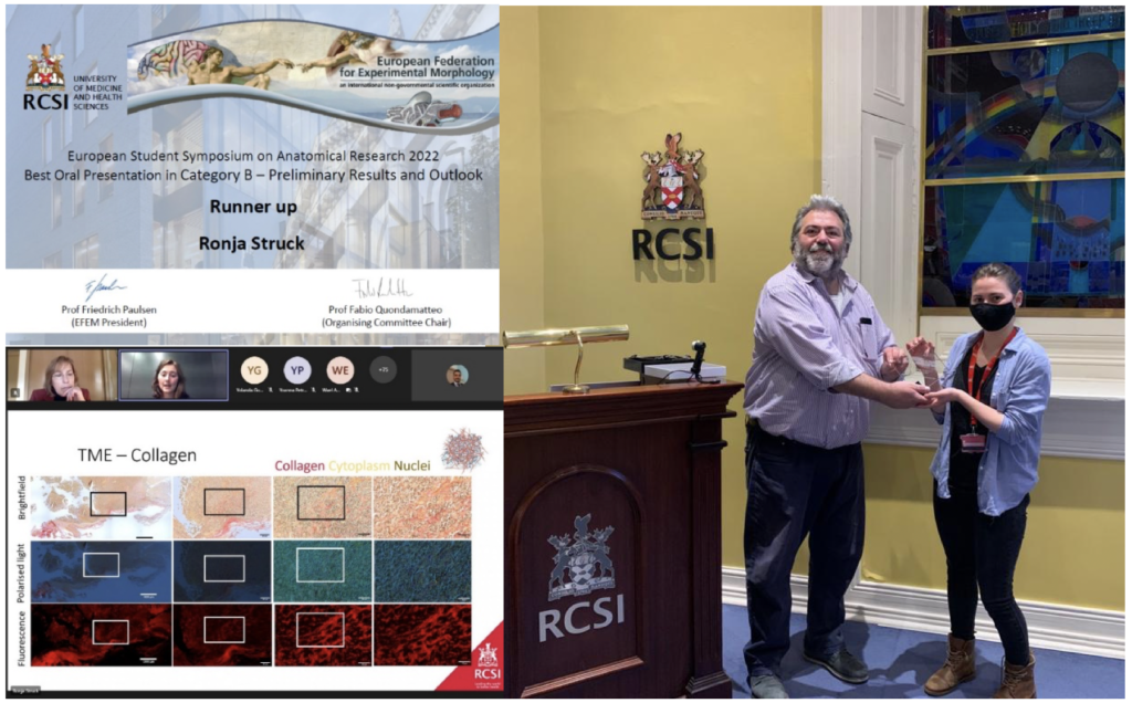
But by far the reason why I’ll remember this conference for the longest time is that being awarded runner up in the category Preliminary Results and Outlook reassured me that I am on the right track and that there is purpose in what I do. Despite the online format of the conference, I had the honour of receiving my prize in person, because Prof. Fabio Quondamatteo, the organiser of the event is based at RCSI.

Overall, the two days were an important step in consolidating my faith in my work and the career path that I have chosen.

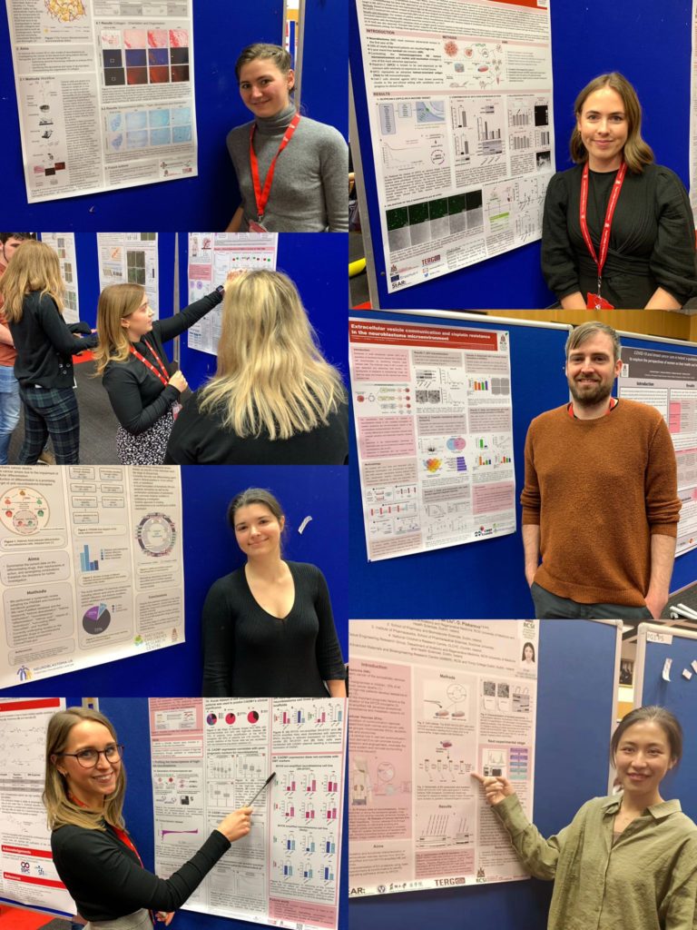
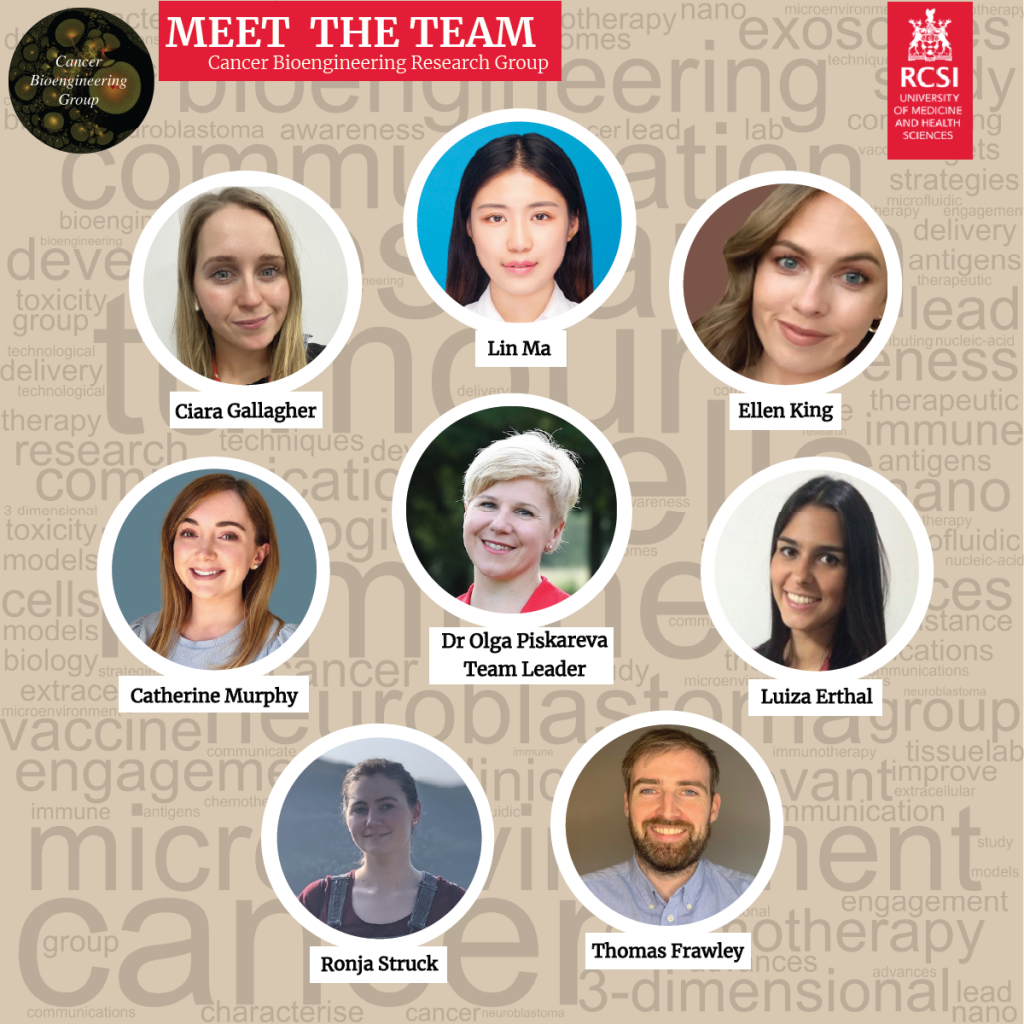
Written by Ronja Struck, the IRC-CFNCRF funded PhD student

RCSI Research Day 2022

Cancer Bioengineering Group thoroughly enjoyed getting back to in-person Research Day at RCSI after 2 years, we’re now very much looking forward to the IACR conference later this month! We will have 2 oral and 5 poster presentations at IACR 2022.

Dream Team in action

Ronja: My typical day

A typical day for me is difficult to describe because there are many facets to a PhD in the Cancer Bioengineering research group. Some days I spend in the lab sectioning, staining or looking at tumour samples under the microscope. Others I stay at home, read papers and try to figure out how they can help me to achieve my research goals. Some days I take part in the courses and workshops offered in the scope of a structural PhD. Then there are times when I sit here writing up for you guys what it is that I do those other days. The academic environment also provides lots of other opportunities to apply yourself and broaden your horizons or pursue what you enjoy. I, for example, have the chance to partake in weekly dissections for medical teaching which helps to keep my anatomical knowledge fresh and is an always welcome change of scenery (and smell) when I am stuck on other things. Furthermore, I get to see the other side of conferences and what is involved in their planning, because I am part of the local organising committee for the European Federation for Experimental Morphology Symposium 2022.

Figure 1 Working on neuroblastoma cancer the samples I am working with are quite unsurprisingly tumour cells. But these can be grown, for example, in mice (A) or on manmade scaffolds (D).  I am moving a staining rack that holds the microscopy slides through staining containers filled with different solutions (C) to stain the slides. After the slides are stained the excess stain is removed by washing in distilled water (B). The resulting images depend on the type of stain. Stains like Alcian Blue can only be viewed with brightfield microscopy (A). But Picrosirius red can also be viewed under polarised light or as seen here (D) with fluorescent microscopy.

Currently, not yet half a year into my PhD, a lot of my time is spent planning. That’s planning which methods to use, which products to order and which experiments, and analyses would result in the most coherent and rounded off story being told by the summation of my research. I also spend a lot of time optimising the methods I will use to assure reproducibility and avoid issues during the analysis later on. For example, the whole tumour sample stained with Alcian blue you can see in Figure 1A clearly shows discernible blue and red regions. However, I have spent about 2 months now trying to get to a point of producing this same outcome reliably rather than having samples show up entirely blue or very only faintly stained. Picrosirius red, the solution I used to stain the sample in Figure 1D stains collagen. But there are many different stains for collagen. After researching most if not all of them I chose this one because it can be viewed with different types of microscopies providing slightly different information. Another step of planning includes how many pictures of which magnification will be required, one image of a whole section for orientation such as in Figure 1A and then more zoomed-in images to investigate the structure of collagen such as in Figure 1D.

Between course work and planning and optimising different aspects of my project, my PhD provides me with plenty of opportunities to focus on something else whenever I get stuck to later return with a fresh set of eyes.

Written by Ronja Struck, a 1st Yr PhD student funded by the IRC-CFNCRF

Welcome to the Cancer Bioengineering Group!

It is time for a full group presentation here at the blog! Throughout the month we shared about our group members and their research focus on Twitter. Now, we would like to share more about the group here and invite you to keep following us on social media. 

The Cancer BioEngineering Group is a research group led by Dr Olga Piskareva at the Royal College of Surgeons in Ireland. The group has 6 PhD students developing research projects around neuroblastoma biology.  

Our projects address topics related to neuroblastoma microenvironment, cell interactions, tumour resistance and the development of new therapies. To do that we use 3D in vitro models, identify immunotherapeutic targets and evaluate extracellular vesicles.  

We are a dynamic group proud to be engaged in research, science communication and patient involvement. We do that through different initiatives.  

We support and collaborate with several neuroblastoma charities around Ireland and internationally such as the Conor Foley Neuroblastoma Foundation, the National Children Research Centre, the Children’s Health Foundation Crumlin and the Neuroblastoma UK. Moreover, our projects are funded by the Irish Research Council in partnership with these charities and by RCSI StAR PhD programmes.  

We promote neuroblastoma awareness through different activities. For instance, last September at the Childhood Cancer Awareness month we promoted a hiking challenge to raise money and increase awareness of neuroblastoma. We hiked for 30km at Wicklow mountains in a day and raised over € 2,000 for neuroblastoma research charities.  

We are also present in social media, creating content in the form of blog posts and tweets to share the science we are doing.  

We are always happy to answer questions and interact with the public. Follow us on our social media channels and read our blog to know more about us and our research.  

Thanks for reading and we go ahead with neuroblastoma research! 

Written by Luiza Erthal

Models to study neuroblastoma in the laboratory

Finding suitable research models to study disease is a big challenge for researchers around the world. In cancer research, it is essential to work with models that can recapitulate tumour characteristics as much as possible. This is important to test chemotherapeutic drugs, understand tumour behaviour and have higher chances of translating the finds from the laboratory to clinical practice.  

Multiple factors influence tumour behaviour and disease progression. The most important is the tumour microenvironment, which comprises different cells and molecules that surround the tumour and the extracellular matrix, a network of molecules that provides support to the cells in the body.  

Most cell studies in a laboratory are based on 2D cell culture models in which the cells grow in a monolayer. Although this approach has a low cost and it is easy to use, it lacks the complexity observed in the clinical scenario. It is true that no model can recapitulate all the complexity found in the body. However, scientists were able to develop interesting approaches to study different tumour characteristics with relatively good approximation1.  

Specifically for neuroblastoma, the most common solid tumour that affects children, scientists developed 3D models in which neuroblastoma cells grow interacting with the surrounding environment and with each other in a vial. Examples of 3D models include cells grown in hydrogels or scaffolds and multicellular tumour spheroids (see image below). Spheroids are formed through the self-adhesion of tumour cells growing in the form of very small balls. They can be maintained in the laboratory on their own or supported by scaffold-based platforms (jelly-like or porous materials). Scaffolds essentially support the cell resembling the extracellular matrix and surrounding tissue in the body. 

In the Cancer Bioengineering Research Group, we work with neuroblastoma models such as organoids, a more complex type of spheroid, to understand neuroblastoma migration and invasion2. Moreover, we recently shared with the research community a protocol at jove.com describing the development of a 3D neuroblastoma model using collagen-based scaffolds3.  

Time-lapse video of neuroblastoma organoids’ growth. Accompanying experimental data published in Gavin et al., Cancers 2021. Source: the Cancer Bioengineering Research Group 

These models have the potential to advance drug tests performed in the laboratory providing better clinical translation, ultimately contributing to improving the quality of life and survival of children diagnosed with neuroblastoma.  

The work with 3D models at the Cancer Bioengineering Research Group is supported by the Irish Research Council, the Conor Foley Neuroblastoma Cancer Research Foundation, Neuroblastoma UK and National Children’s Research Centre. 

Written by Luiza Erthal

References 

1. Nolan, J. C. et al. Preclinical models for neuroblastoma: Advances and challenges. Cancer Lett. 474, 53–62 (2020). 

2. Gavin, C. et al. Neuroblastoma Invasion Strategies Are Regulated by the Extracellular Matrix. Cancers 13, 736 (2021). 

3. Gallagher, C., Murphy, C., O’Brien, F. J. & Piskareva, O. Three-dimensional In Vitro Biomimetic Model of Neuroblastoma using Collagen-based Scaffolds. J. Vis. Exp. 62627 (2021) doi:10.3791/62627.