A new 3D strategy to study neuroblastoma

Our body has 3 dimensions: height, width and depth. Every single part of our body grows in the same 3 dimensions. This is true for cancer cells. Researchers use different ways to study cancer cells behaviour, how they grow and spread. We grow cells in the flasks, where they change their structure and shape and become flat losing one dimension. This is a very popular approach. We also grow cells in mice, where cells keep their 3D shape and mimic their behaviour to one observed in humans.

It is well known that we need to give a different amount of drug to kill cancer cells grown in flasks and in mice. This, in turn, delays the development of new drugs. Why does it happen this way? So, the drug works only on one side of the cell when they grow on the flat surface. In contrast, in mice, drug surrounds the cancer cell habitat and attacks cells at the edge first and then getting to those at the core. So we need more drug to kill cancer cells in mice.

We decided to design a new way to grow cancer cells that recreate their growth in 3 dimensions as in the human or mice body. We used special cotton wool like sponges as a new home for cancer cells and populated them with cancer cells. At the next step, we gave cells the drug at the different amount and checked what happened.

To understand cell fitness we stained them with red and blue dyes. On the left bottom side of the image, we see an equal amount of red and blue dyes telling us that cells were healthy and fit. Cells did not get any drug. When we gave a little amount of the drug but enough to kill cells in the flask, the balance of red and blue dyes was the same telling us that nothing really happened (the image in the middle). Cells were feeling well and healthy. The right bottom image has only blue dye. In this case, cells were given the amount of drug enough to destroy cancer cells in mice or humans. The lack of red dye tells us that this time the drug worked and killed the cancer cells.
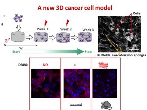
We found that the drug killed cells on sponges only at doses enough to do the same in mice.

So, we concluded the new tactic to grow cancer cells in 3D on cotton-like sponges can bridge the gap between traditional way and animal models. This new strategy to grow cells on sponges should help to understand cancer cell behaviour better and accelerate the discovery and development of new effective drugs for neuroblastoma and other cancers. This, in turn, will make the outlook for little patients better and improve their quality of life.

This work has been published in Acta Biomaterialia and presented recently at the Oral Posters Session at the 54th Irish Association for Cancer Research Conference 2018.

This study was supported by Neuroblastoma UK and National Children’s Research Centre.

You can find more at
A physiologically relevant 3D collagen-based scaffold–neuroblastoma cell system exhibits chemosensitivity similar to orthotopic xenograft models.

IACR Meeting 2018 Programme

Irish Neuroblastoma Research Collaboration

On November 20th, the Irish neuroblastoma researchers have met for the first time to set up a collaborative research hub.  The aim is to consolidate their expertise and skills in order to crack the neuroblastoma code together.

They all have different science background spanning from molecular and cellular biologists,  immunologists, tissue-engineering, bioinformatics, mathematical modelling and clinicians representing RCSI, UCD, TCD, OLCHC and NCRC. During this meeting, researchers talked about their challenges and progress finding out that we are complementing each other projects. Clinicians from different OLCHC departments exposed basic researchers to realities of the disease.  None would find this information in academic papers: it is what you see in the clinic and how it works in practice.

Big thank you to Dr Cormac Owens for the invitation and linking us together and Prof Jacinta Kelly for mapping the support available from the NCRC and CMRF.

Our next meeting will be held in RCSI in January 2018.

Happy Birthday the Irish Neuroblastoma Research Consortium!


What lessons have been learnt?

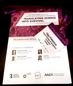
Today is the final day of the Third International Cancer lmmunotherapy Conference. The meeting was run at the Rheingoldhalle Congress Center in Mainz/Frankfurt, Germany from September 6-9, 2017. More than 500 people attended this meeting.

The focus of the scientific program was on “Translating Science into Survival”. Talks covered the challenging areas in cancer immunology and immunotherapy. The full list of topics can be found in the meeting program.

At the moment cancer immunology and immunotherapy is a hot topic in the next generation of anti-cancer therapies. Lots of attention is given to checkpoint immunodrugs as it was proven by the prevalence of talks on this subject in the program. Indeed, this drug has great potential, but at the same time, it is not universal. About 50% of patients do not benefit from it.

What lessons have been learned from the talks:

  • Checkpoint immunotherapies are the main stream
  • Not all cancer patients would respond to immunodrug
  • Genetic landscape of a tumour and/or the patient may contribute to this, thus making beneficial to check genetics for this type of treatment
  • Immunodrugs work better in combination with conventional therapies such as chemotherapy.
  • The immune system can be tuned by a drug, but it will switch on compensatory mechanisms to balance the intervention.
  • Lots have to be studied further


Father of Chemotherapy and Cancer Immunology

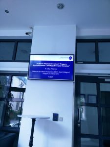
I was giving a talk at Georg-Speyer-Haus Institute for Tumour Biology and Experimental Therapy yesterday. The aim of my visit was to establish collaboration with Prof Daniela Krause, who is the expert in bone marrow microenvironment and targeted therapies. She took me to the Institute museum that keeps the history of this place and phenomenal researchers used to work there.

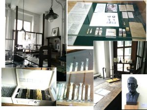
This research institute was established in 1904 to support work of Paul Ehrlich, its first director and funded by the private foundation “Chemotherapeutisches Forschungsinstitut Georg-Speyer-Haus”. Paul Erlich is the Father of the chemotherapy concept originally developed to treat diseases of bacterial origin. He reasoned that there should be a chemical compound that can specifically target bacteria and stop its growth. He developed Salvarsan, the most effective drug for treatment of syphilis until penicillin came onto the market.

Paul Erlich is also known for his contribution to cancer research. He and his colleagues actively experimented on how tumour originates and spread. They also tried to understand how immune system can beat cancer applying vaccination concepts.

Paul Erlich’s Lab back then. Now it is a museum

Paul Erlich and Ilya Mechnikov were jointly awarded The Nobel Prize in Physiology or Medicine for his “work on immunity” in 1908.


The Nobel Prize Diploma

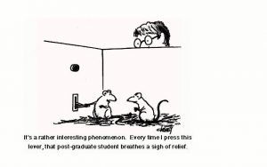
August is a very quiet month

It is very quiet in the lab this month. No troubleshooting, no more long working hours, endless repetition of experiments, smiles and upsets… Almost all students completed their projects, submitted their works for grading and graduated. The last student is finishing at the end of August.

Time to focus on the collected data, reading literature, writing papers and new grants.

http://www.ifunny.com/pictures/its-rather-interesting-phenomenon-every-time-i/

Research Summer School in Action

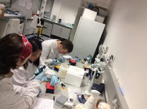
It is always a pleasure to host undergraduate students during summer months. Two students joined the RCSI Research Summer School (RSS) Programme. Both are working on the NCRC funded project to understand mechanisms that drive neuroblastoma pathogenesis. None of them had a prior lab experience, but nothing is impossible under John’s supervision.

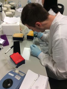
A full concentration on every single step of the research.

Congratulations to Dr.John Nolan!

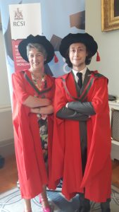
My PhD student John Nolan together with other 41 candidates graduated at the RCSI’s 2017 June Conferring ceremony which took place in the College Hall of 123 St. Stephen’s Green.

He continues his research in neuroblastoma as a Postdoctoral researcher on the project funded by the National Children’s Research Centre. I am glad to be able to keep expertise and young talents in our team.

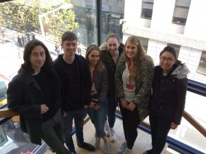
Neuroblastoma Research Dream Team 2017

It is fantastic to see so knowledgeable and enthusiastic young researchers in my research group. This year, the team is multinational with the Irish students mixing with Belgian and Malaysian. All together they are cracking the code of neuroblastoma microenvironment and tumour cells communication through understanding main differences between conventional cancer cell models and tumours.

The big research plan of the entire team consists of more smaller and focused projects to be completed within 10-12 weeks. All projects are unrestricted, they are driven by the intellectual curiosity of these students. This way is full of ups and downs, frustrations and encouragements when techniques do not work or reagents do not come in as expected. Some cancer concepts can also work differently in the given settings. Simple questions are bringing more challenges than expected.  But at the end of the road is the best reward – contribution to the conceptual advancement of neuroblastoma microenvironment.


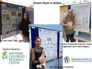
The Neuroblastoma Research Dream Team 2017: Dr. John Nolan, NCRC funded researcher, RCSI, Joe O’Brien, TCD MSc student, Ciara Gallagher, DIT undergraduate student, Jessica Tate, RCSI Medical student, Larissa Deneweth, Erasmus student, Ghent, Ying Jie Tan, TCD MSc student.

Identification of biomarkers of tumour response to drugs

A Research Project Grant funded by National Children’s Research Centre will be starting in April.

The ultimate aim is to identify biomarkers of tumour response to drugs in the blood of children with high-risk neuroblastoma.

Challenge: Treatment regimens for patients with high-risk neuroblastoma involve intensive, multi-modal chemotherapy. Many patients response to initial therapy very well, but has only short-term effects, with most becoming resistant to treatment and developing progressive disease.

The project has two parts which complement each other.

Part 1

  1. We will study cell-to-cell communication using cell-based models. We will collect exosomes, small envelopes containing bioactive molecules, produced by drug-resistant cell lines to treat non-cancerous cells. We will measure the effect of exosomes on non-cancerous cells by counting cell growth, examining their shape and metabolism. We will also examine whether non-cancerous cells treated with exosomes become less responsive to chemo drugs.
  2. We will treat neuroblastoma cells with a drug and collect exosomes before and after treatment. We will profile exosomes to identify any changes in their miRNA content. MiRNA are very small pieces of genetic material that can change the way cell feels and works. This step will help to find biologically active miRNA that can trigger cell resistance to drugs. These biologically active miRNA can represent biomarkers of tumour response to chemotherapy.

Part 2

  1. We will screen clinical samples for exosomal miRNA in response to drug treatment. We are planning to use a small sample of blood taken from neuroblastoma patients during routine examinations before, during and after chemotherapy.This step will help to find clinically relevant miRNA of tumour responsiveness to chemo drugs.
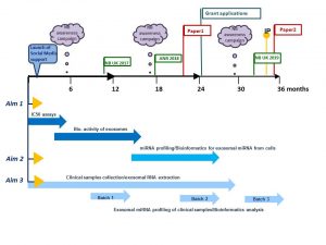
The plans for 3 years

How does this project contribute to the biomedical community?

This study aims to contribute to the better understanding of the disease mechanisms and scientific knowledge in the area, and in particular how neuroblastoma cells communicate with other cells helping tumour to create a unique microenvironment and protect themselves from chemotherapy pressure. The new data will give insights in biologically active proteins and miRNAs involved in cell-to-cell communication and drug responsiveness.

What are potential benefits of the proposed research to neuroblastoma patients?

This project aims to develop exosomal biomarkers of tumour response to drugs that might be used to help select patients for treatment and identify novel targets for the development of more effective personalised therapy with the anticipated improvement in outcomes. This work will contribute to the more efficient design of re-initiation treatment, sparing patients unnecessary rounds of chemotherapy and ultimately increasing survival. These new circulating markers will benefit children with high-risk neuroblastoma whose tumours are relapsed leading to less harmful and more tailored treatment options and improving their quality of life.