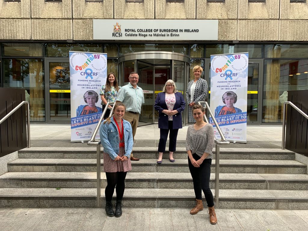
We will work closely with the Conor Foley Neuroblastoma Cancer Research Foundation – a research charity led by the family who lost their child to neuroblastoma. An inspirational example of never giving up.
We will continue to dissect neuroblastoma biology using innovative platforms such as tumour-on-chip and 3D scaffold-based models in collaboration with our colleagues in the Tissue Engineering Research Group at RCSI and the Fraunhofer Project Centre at DCU.
This announcement is timely to celebrate Childhood Cancer Awareness Month in September.
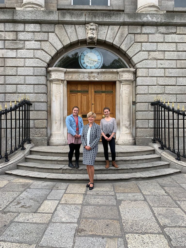
Two talented and dedicated young scientists are joining our team. In 4 years time, we will have another pic of their graduation on the same stairs.
Regardless COVID19 pandemic, we continue to host undergraduate students from various Universities for their research projects. Two students, Carla and Chris, from the Technical University of Dublin, carried out BSc projects remotely. Having in-house datasets and many more published in open access, their projects were focused on bioinformatics, re-analysing them and giving a second look. Both Carla’s and Chris’ research received the highest score in their classes. Many congratulations – well deserved!! We wish to thank both for their kind words and willingness to share their story.
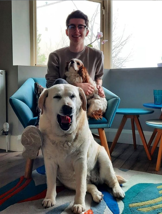
Chris Sheridan, the final year student in Biomolecular Science at the Technical University of Dublin, 2021
“My project concerned analysing the exosomal miRNA expression of neuroblastoma cells in response to chemotherapy. Though the project was not too large, it certainly was the largest project I have ever taken part in. The work Dr. Piskareva and her team are conducting is so interesting and novel that I felt very fortunate to be participating in such an exciting field. Despite the novel and complex nature of the topic, the project was extremely engaging, allowing for an opportunity to learn new valuable research and data analysis skills. I was able to get very useful and helpful feedback regularly from everyone on the research team, where there was a very welcoming and positive attitude. This made the topic seem less daunting and my goals more achievable. I was really happy with my results, and I am excited to see where they may lead in the future. Some of the miRNAs identified in the analysis may represent potential biomarkers or therapeutic targets for high-risk neuroblastoma patients. As I have yet to experience any lab-based research, it was cool to see the team’s approaches and applications of lab techniques and analysis strategies to see how research is conducted in the “Real World” after seeing these topics before only in lecture notes. Overall, the project was challenging but very rewarding and enjoyable. Throughout the project, the overall experience, the excitement of the results coming together, and the realisation that I may have something to contribute to this field of research cemented the idea in me that this is certainly the path I wish to pursue in science and for that, I would like to thank Dr. Piskareva and her team for such a positive and educational experience during my time with them.“
Carla Tejeda Monné, the final year Technological University of Dublin Biomolecular Science Student, specialising in Biotechnology, Therapeutics, and Drug Development, 2021
“During my final year project, I had the unique and amazing opportunity to work under the supervision of Dr. Olga Piskareva. The purpose of my thesis was to assess the clinical significance of Tumour Necrosis Factor Receptor Superfamily Member 1B and Member 4 (TNFRSF1B and TNFRSF4) in neuroblastoma patients. I accomplished this by analysing the gene profiles of several tumours using bioinformatic tools. In addition, I investigated the potential of microRNAs as therapeutic agents for neuroblastoma treatment. I thoroughly enjoyed carrying out this research project, and I hope the findings from my thesis can aid future research into the pathogenesis of neuroblastoma and the development of effective treatments for these children.“
Best of luck to Chris and Carla in their next endeavour!
Yep, we are living in challenging and extraordinary times. The COVID19 changes and dictates rules, but training of future health professionals is going on.
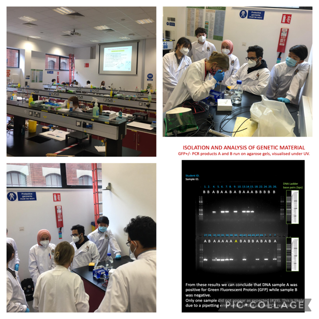
Within a fantastic RCSI summer training programme for medical students, our team ran essential practicals on the isolation of genetic material and the use of polymerase chain reaction, known as PCR, to detect differences in normal and modified genomic DNA.
Polymerase Chain Reaction, or simply PCR, was conceived and validated by biochemist Kary Mullis in 1983. This discovery revolutionised many scientific fields that dealt with genetic material and was awarded a Nobel Prize in Chemistry in 1993. PCR allows rapid generation of small identical fragments of DNA. The fragments can be visualised, their size and number can be calculated. It has become a standard procedure in molecular biology and pathobiology screening. The COVID19 PCR test is actually an advanced modification of Mullis’ invention.
All students successfully set up individual PCRs to our great satisfaction, and the results are presented at the right bottom corner.
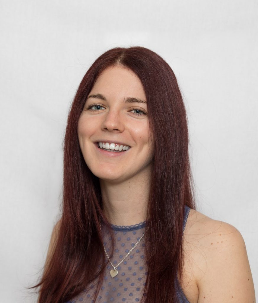
In early April, shortly after attending the Irish Association for Cancer Research (IACR) virtual conference, I was delighted to find out I had been selected for a “Poster in the Spotlight” presentation at the European congress in June. I was invited to give a 10-minute presentation about my research on finding new immuno markers for drug-resistant neuroblastoma. This would be my first time presenting at a conference outside of Ireland (though I remained in my home in Dublin and not in the hoped destination of Turino) and so it was a very exciting experience!
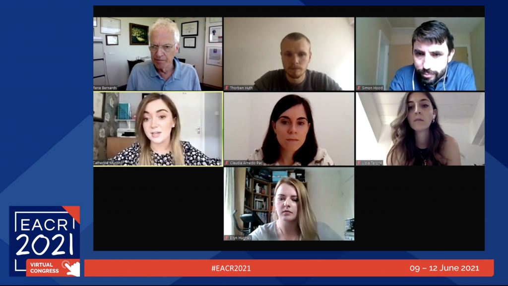
On the morning of my talk, I logged onto zoom to meet the EACR session organisers and the other speakers in my session – a mixture of PhD students and Post-doc researchers from Germany, Spain, Italy and the UK. Each of our talks was then broadcasted to the virtual congress platform where cancer researchers around Europe could tune in.
After the presentations, we joined a “Meet the Speakers” session where we could chat with those who had tuned in, answer questions and open up some research discussions. I was asked one question from a researcher doing similar work to me on how I investigate the relationships between certain genes in cancer, and I was able to refer her onto the software I use – I hope this aids her research, as that’s the real aim of attending these conferences!
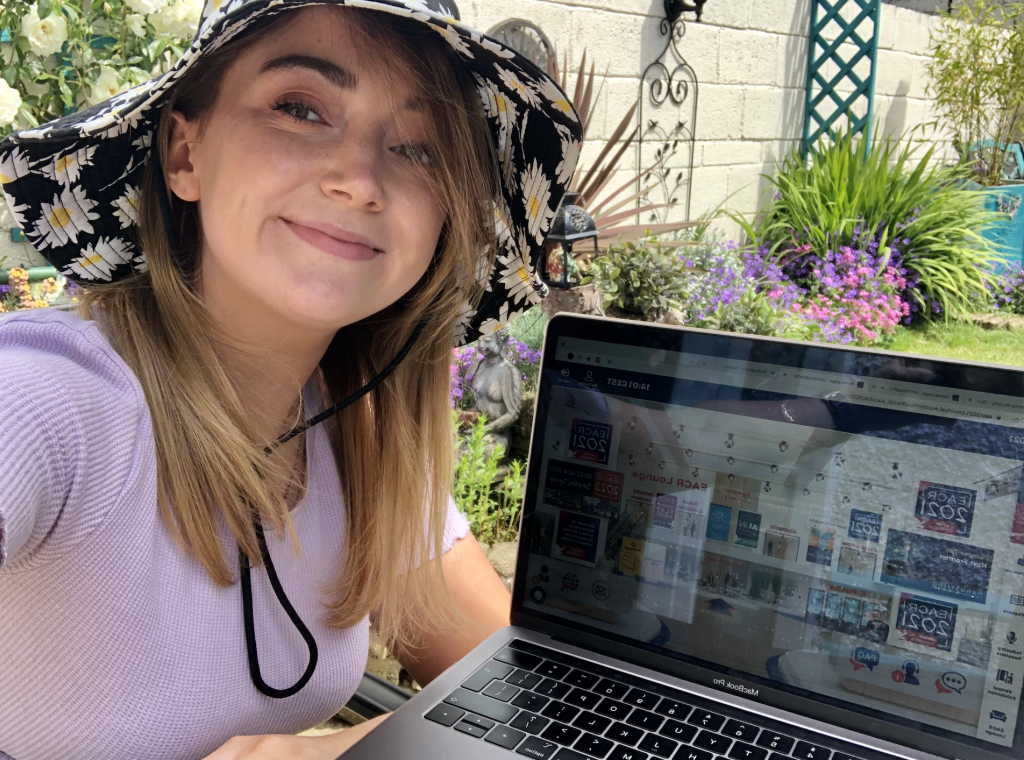
Following this, it was time to relax and watch some of the other interesting talks taking place in the congress. The EACR were running a photo competition to show where you were watching from in order to win a place at the 2022 congress in Seville. So, I took this opportunity to take my laptop out to the back garden and watch the congress in the sun, getting a selfie for twitter with #NotQuiteTorinoEACR2021. Fingers crossed by this time next year travelling for international conferences will be a reality for researchers once again!
Catherine Murphy, Neuroblastoma UK funded PhD student
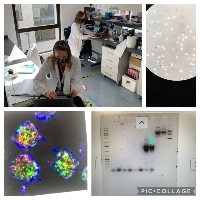
Sunny and chill outside. But PhD students are very busy in the lab with lots to do, record and analyse. Happy with small steps forward. The right size of a PCR band (bottom right), nice looking recombinant bacteria colonies (top right), neuroblastoma cells formed perfect spheroids (multicolour image).
Once I mentioned the importance of the publication track record for a career in science. My team has been productive despite the COVID pandemic. Two review articles were published.
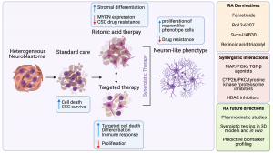
The second review has been published in Journal of Personalized Medicine on March, 16th. Originally, it was a small review project for Nadiya, a medicine student, last summer. However, it became a big one with all data systematically collected, analysed and condensed. The focus of this review was on Retinoic Acid (RA), widely known as Vitamin A and its role in neuroblastoma. RA plays a vital role in human development. The main feature of RA is to push neuroblastoma cells to become neuron-like cell stopping their aggressiveness and cancer fate. So, we wanted to know more about the ongoing research both in the labs and the clinic. We reviewed primary research articles reporting basic and translational findings as well as clinical trials. Hopefully, it would help other researchers to get a full picture of this topic and a structured resource of experimental models and drugs tested.
Since I joined neuroblastoma research, I have been puzzled by the fact that half of the children with neuroblastoma have the disease spread at the time of diagnosis. It is still a puzzle whether cells spread and primary tumour growth happen simultaneously or more adventurous cancer cells escape the primary tumour location later.
At a cancer conference, I met Prof Ewald who studies this process in breast cancer. I was fascinated by the approach and started to look for opportunities to join his lab. To tell the truth, very few exist for mid-stage career scientists! One of them is the Fulbright program.
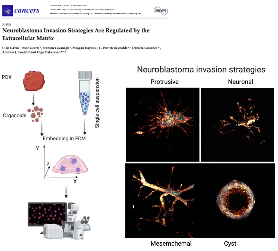
One day, I opened my email saying that I received a Fulbright-HRB Health Impact Scholar Award to travel to Johns Hopkins University and adapt their 3D models to learn how neuroblastoma spreads. It was a life-changing experience both personally and professionally. The amount of experimental data collected over 4 months of work did not fit a 1TB memory stick! Indeed, this short journey was just a start of a new research inquiry.
On my return home, the greatest task that remained was to make sense of every single experiment. Cian Gavin took over and spent almost a year systematising, characterising it, and placing it into a context. It was meticulous work with very little known about invasion strategies in neuroblastoma. Now, we are happy to share our findings published on Cancers.
Where do we go now? Well, our next step is to understand the cellular players behind neuroblastoma invasion and how we can target them to stop neuroblastoma spread. It won’t be a short and sweet journey, but we are ready for it!
This fantastic and rewarding work was supported by Fulbright Commission Ireland, National Children’s Research Centre, Health Research Board, Science Foundation Ireland, the National Institutes of Health/National Cancer Institute (Prof Ewald), Alex’s Lemonade Stand Foundation for the COG Childhood Cancer Repository (Prof Reynolds) and the National Institutes of Health/National Cancer Institute (Prof Reynolds).
So, the 2021 has begun and the COVID-19 is still challenging us.
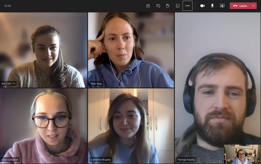
Our first lab meeting this year was on Weds, and our, now traditional, coffee morning has happened today. It usually happens on Fridays.
What did we chat about for almost an hour? Well, about making favourite pet’s drawing as Christmas gifts, the development of bicycle’s and bus’ infrastructure, baking recipes, new life targets, like reading more books and doing more healthy stuff.
Research is a fascinating journey no doubt. Inquisitive minds try to solve burning puzzles. It takes time. Some puzzles are more complected than the others. One of the hallmarks is the conversion of the resolved puzzle into a scientific story to tell to your peers.
We write and publish these stories. The publishing is another caveat that often makes your story sharper and neater. However, while you are in the process you feel that the mission is impossible.
Delighted to see that one of the missions is completed – a great hallmark for John which coincided with his new research adventure starting in a few days. This is his first first author paper! It is not tautology! It is his first original research paper where he is the first author. This position is a success measure in a research career. His teamwork skills secured him another few original papers. Well done John! Well deserved!
Last month we set ourselves the “10 Laps 10km” challenge for Childhood Cancer Awareness.
Now we have closed the GoFundMe and counted the charity buckets. We are delighted to announce we raised a grand total of €1419! We are over the moon with this sum, as 2020 required a very different kind of fundraiser than previous years.
Our three chosen charities: Children’s Health Foundation Crumlin (formerly CMRF), the Conor Foley Neuroblastoma Cancer Research Foundation, and Neuroblastoma UK, will each receive just over €470.
We’d like to say big thank you to everyone who donated. It will make a huge difference for these charities, this year especially, paving the way to better treatment options for children with cancer in the future.