Towards 3D neuroblastoma cell models

It seems I have got a conference season. Three conferences within 2 months – no complains though. This time I went to the Matrix Biology Ireland Meeting in Galway. It was fantastic mix of topics and speakers ranging from new approaches in bone and heart repair to new matrixes in reconstruction of body tissues and diseases in the lab to minimise use of animals in pre-clinical studies.

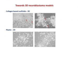
My talk was focused on neuroblastoma microenvironment and cell-to-cell communication through exosomes. I wrote about it in October post. I talked about things that did work and did not as well as new directions. One of the new directions is reconstructing neuroblastoma by growing neuroblastoma cells on collagen based scaffolds in 3D. Collagen constitutes most of our tissues to keep it shape and strength. These scaffolds are sponge-like matrixes built from collagen and other components. Of course cells grow differently on these matrixes. They have a different shape and growing properties in 3D. Neuroblatoma cells look like water drops on the cotton wool-like collagen scaffolds. In contrast, when they grow on plastic in 2D, they are flat. Studies show that cells in 3D respond to cytotoxic stress in a similar pattern as if being within a body (details in recent reviews 1-4).  It would be a great breakthrough once these models are optimised for neuroblstoma research  field. It will help to test all new and known drugs in the environment close to clinical settings. It could be a step forward to personilised therapies for children with neuroblastoma by isolating cancer cells, growing them in 3D and testing how they respond to all therapies available. It will facilitate more efficient design of treatment for relapsed or poorly responding tumours, sparing patients unnecessary rounds of chemotherapy and ultimately increasing survival.


Neuroblatoma cells look like water drops on the cotton wool like collagen scaffolds. In contrast, when they grow on plastic in 2D, they are flat. Arrows point towards cells.
This is microscopic images of neuroblatoma cells growing on the collagen scaffolds and plastic. Arrows point towards cells.


I’ve always felt that a selection of abstracts for an oral presentation is biased. The overall background and views of conference organisers would affect works selected for an oral presentation.  The same abstract was not selected for an oral presentation by one committee, but was supported by the other.  Never give up!

Readings:

  1. Schweiger PJ, Jensen KB.Modeling human disease using organotypic cultures. Curr Opin Cell Biol. 2016 43:22-29.
  2. Salamanna F, Contartese D, Maglio M, Fini M. A systematic review on in vitro 3D bone metastases models: A new horizon to recapitulate the native clinical scenario? Oncotarget. 2016 7(28):44803-44820.
  3. Picollet-D’hahan N, Dolega ME, Liguori L, Marquette C, Le Gac S, Gidrol X, Martin DK. A 3D Toolbox to Enhance Physiological Relevance of Human Tissue Models. Trends Biotechnol. 2016 34(9):757-69.
  4. Nyga A, Neves J, Stamati K, Loizidou M, Emberton M, Cheema U. The next level of 3D tumour models: immunocompetence. Drug Discov Today. 2016 21(9):1421-8.

Quality of life for childhood cancer survivors

For children who do survive cancer, the battle is rarely over.  Over 60% of long‐term childhood cancer survivors have a chronic illness as a consequence of the treatment they received; over 25% have a severe or life‐ threatening illness. How much do we know about quality of life of childhood cancer survivors?

Researchers in health- and illness-related social sciences understand that the there is a life after the treatment completed. The life is full if diverse levels and issues from health related to social adaptation in different shapes and forms. Children and teenagers may experience fear when returning to school due to temporary or permanent changes to their physical appearance (1,2). They worry about their ability to socialise with their friends due to lengthy absences (3–5). Treatment can result in the development of learning disabilities in children and thus marking school as a major source of frustration (1,2). These learning difficulties can affect a child’s confidence and self-esteem, if left without attention and care (1,3). All studies come to the same conclusion. Challenges in education of children with cancer are complex, however most can be tackled efficiently through planning and good communication (1–5).

Recently researchers working in FRED HUTCH Cancer Research Center asked adult childhood cancer survivors a number of health related questions about the quality of lives (6,7). The results are far from optimistic: “chance of surviving childhood cancer has improved — but survivors’ overall health has not”. You can find more by following the link.

It is important not only to recognise the problems but to start changing the situation. Apparently much more could be done more efficiently if patients are involved in setting up future research agenda.

Reading

  1. Gurney JG, Krull KR, Kadan-Lottick N, Nicholson HS, Nathan PC, Zebrack B, et al. Social outcomes in the childhood cancer survivor study cohort. J Clin Oncol. 2009;27(14):2390–5.
  2. McDougall J, Tsonis M. Quality of life in survivors of childhood cancer: A systematic review of the literature (2001-2008). Supportive Care in Cancer. 2009. p. 1231–46.
  3. Barrera M, Shaw AK, Speechley KN, Maunsell E, Pogany L. Educational and social late effects of childhood cancer and related clinical, personal and familial characteristics. Cancer. 2005;104(8):1751–60.
  4. Langeveld NE, Stam H, Grootenhuis MA, Last BF. Quality of life in young adult survivors of childhood cancer. Support Care Cancer. 2002;10(8):579–600.
  5. Klassen AF, Anthony SJ, Khan A, Sung L, Klaassen R. Identifying determinants of quality of life of children with cancer and childhood cancer survivors: A systematic review. Support Care Cancer. 2011;19(9):1275–87.
  6. Yeh JM, Hanmer J, Ward ZJ, Leisenring WM, Armstrong GT, Hudson MM, et al. Chronic Conditions and Utility-Based Health-Related Quality of Life in Adult Childhood Cancer Survivors. J Natl Cancer Inst [Internet]. 2016;108(9):4–7.
  7. Armstrong GT, Chen Y, Yasui Y, Leisenring W, Gibson TM, Mertens AC, et al. Reduction in Late Mortality among 5-Year Survivors of Childhood Cancer. N Engl J Med. 2016;374(9):833–42.

E-poster at SIOP2016

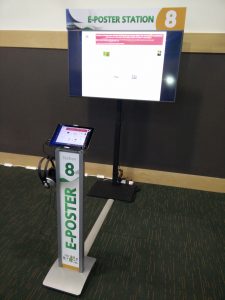
Ok. Now, when the stress of the presentation is over, I am happy to share new technologies used during the SIOP2016. As I mentioned yesterday, my work was selected for e-poster presentation. It looked this way:

This is e-poster station, where anyone can look up all posters displayed during the meeting.


It is definitely a step forward. Anyone can look up any poster, listen to a commentary recorded by the author, zoom in and out and send a request/comment to the author. It looks cool and trendy. Though, you can feel invisible as no physical copy displayed in a designated area. No crowds of poster presenters and judges. No waiting faces desperate to share their study…

The actual Poster Discussion session was a traditional presentation when my poster was up on the big screen, I had 8 minutes to convince the audience navigating through figures. This session was late and no many attendees survived to come and challenge your statements. Nevertheless, it was enjoyable experience. 🙂

siop-2016-banner-728-x-90


Last minute discovery with SIOP2016


SIOP is the International Society of Paediatric Oncology. It is a global multidisciplinary society representing doctors, nurses, other health care professionals, scientists and patients or their relatives. The Society’s motto is ‘no child should die of cancer’. The meeting 2017 is being held in Dublin, the city where I live and work.

Indeed, it was appealing to attend the key meeting in childhood oncology field. As any participant, I had an opportunity to submit an abstract about my research.  To no surprise at all, I received email notifying me on my work being selected for e-Poster presentation.  Common stuff. The email also said that it would be displayed at designated stations, like big screens throughout the meeting. Very unusual format, but we are living in the digital technology era; things are changing all the time. So, I would not need to stay by the poster this time. Great – more time for networking and talks.

Then I received another email informing about a Poster Discussion session, which I assumed to be a standard procedure when a group of selected piers stand by your poster and ask Qs. None comes in majority cases. A participant stands and waits and waits till the session is over. So, of course I took it easy.

A day before the meeting, I downloaded the meeting app and started to browse along the content and features. Out of curiosity, I checked details of the Poster Discussion session. This was the moment of mental breakdown – I discovered being selected for an oral poster presentation! My chances were 1 in 1475 (the number of submitted abstracts). I should probably also buy a lottery ticket tonight. Could lucky things come together?

I will reflect on the new e-poster presentation experience later today…


siop-2016-ppt


http://www.siop2016.kenes.com/


Newspapers coverage of neuroblastoma: part 1

We do not hear much on neuroblastoma or childhood cancer in our everyday life unless we know a child affected by the disease. How much information can we get from newspapers? How do newspapers report it? What is their focus – a child, his/her family or social circles? In the next posts I will try to get insights from the content of newspaper’s stories.

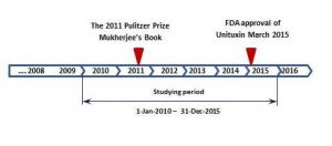
To start with, I needed to select those newspaper articles that cover a story of a child with cancer over a period of time. To do that I selected a recent period  from January 1, 2010 to December 31, 2015 because two major events happened during this time.

The first was the 2011 Pulitzer Prize book written by Siddhartha Mukherjee ‘The Emperor of All Maladies: A Biography of Cancer’ which describing the history of both adult and childhood cancer treatment development and research (Mukherjee 2010).  Importantly, this book inspired to produce a documentary film of three episodes of two hours each and released in 2015 (Goodman 2015). Researchers in social sciences agree that the publication of a prominent popular science book can lead to increased media interest in the issues raised in the book (e.g.  (Nisbet & Fahy 2013).

Another most recent milestone was a breakthrough in childhood cancer management – the FDA approval of a novel drug Unituxin (dinutuximab) for neuroblastoma, the most common solid tumour in children (U.S. Food and Drug Administration 2015).


The timeline of media coverage analysis


next step is actual paper selection. There is an archive of all newspapers and magazines called  Nexis®UK database. This database stores copies of all printed papers and magazines worldwide as well as online versions.

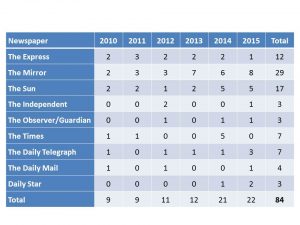
Then an a search for articles carried out looking for key words:‘child’, ‘children’, ‘childhood’, ‘kid(s)’, ‘cancer (s)’, ‘tumour (s)’, ‘treatment’, ‘chemotherapy’, ‘radiation’ and their combinations.  Only articles published by the UK national newspapers and included key words in either the headline or text were selected. The search returned 255 articles. Of 255 articles, 84 contained a story about a child with cancer. The rest were standardised obituaries in the ‘announcements’ or ‘deaths’ sections, horoscopes containing the word ‘cancer’, fundraising and charity activities not involving an identified child, funding initiatives related to pure cancer research and service, reports on scientific achievements or challenges were excluded.

The selected 84 articles were published by the broad range of local and national papers that are daily on sale through news outlets to a general public in the UK.

Summary of selected newspapers


The Q1: What has been the level of media attention to childhood cancer?

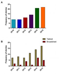
In the last three years, in average 20 articles per year across 9 UK newspapers were published telling us a story about a child with cancer. In other terms it is less than 2 stories per month. It is not enough to raise awareness, educate the public and form their opinion. Tabloid editorial was more interested in this type of stories than broadsheet.  This observation is likely due to the nature of tabloid paper interests looking for people personal stories, entertainment, sports and scandal.

Unfortunately, no change in media coverage of childhood cancer was observed around these two milestones (in 2011 and 2015). All together it suggests that neither of them had a strong influence on journalists or editorials regarding childhood cancer coverage.

Frequency of stories about a child with cancer by year (A) and type of newspaper oulet (B)
Frequency of stories about a child with cancer by year (A) and type of newspaper(B)

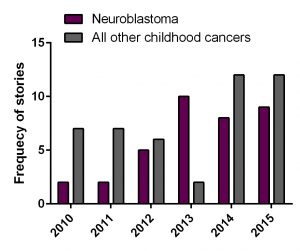
Interestingly, the growing trend in media coverage of children with cancer occurred by the increase in neuroblastoma coverage. Neuroblastoma had a nearly two fold boost in 2013 vs 2012 and then slightly declined in 2014 and 2015. Nevertheless, the FDA approval did not trigger attention to this cancer.


Frequency of stories about a child with cancer by cancer type


Reading

Goodman, B., 2015. Cancer: The Emperor of All Maladies, United States.

Mukherjee, S., 2010. The Emperor of All Maladies: A Biography of Cancer, US: Scribner.

U.S. Food and Drug Administration, 2015. FDA approves first therapy for high-risk neuroblastoma. FDA News Release.

* This study is a part of Dissertation submitted in partial fulfillment for the degree of M.Sc. in Science Communication


Tumour cells travel in a group to new destinations

The conference on models and tumour microenvironment has brought together International experts in this field. Two keynote speakers (Peter Friedl, Radboud UMC/MD Anderson and Andrew Ewald, John Hopkins University) presented exhaustive experimental data on plasticity and microenvironmental control of cancer invasion and metastasis.

Their research teams independently found that

  1. Tumour cells migrate collectively as a team from a piece of tumour like a group of people who changed their minds and decided to travel by bus when the majority stayed camping. However, Andrew Ewald acknowledged that they are not pioneers in this discovery. In 1976 Liotta observed migration of tumour cells in a group of 6-10 cells.
  2. A migration group of cells has their leaders who crave the path through surroundings to the new locations.
  3. Leader cells depend on cancer types. It can be any tumour cell in some cancer types or a specialised one.
  4. Migrating cells take shape and follow the pattern of tissues to be invaded.

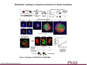
The experiments by Ewald’s research team on collective cell migration. In short, they co-implanted two lung tumour cell populations labelled differently into mice. One cell population had a green protein tag, another had red. After 6-8 weeks, researchers examined metastases and found that they had a mixed population of green and red tumour cells.


Multicellular seeding is a frequent mechanism for distant metastasis. (A) Schema of multicolor lineage tracing assay. ROSAmT/mG;MMTV-PyMT tumor organoids were treated with adenoviral Cre to induce recombination from membrane tdTomato (mTomato) to membrane eGFP (mGFP). Mosaic tumor organoids were then transplanted into nonfluorescent NSG host mice. After 6–8 wk, lungs of these mice were harvested. If metastases arise exclusively from single-cell seeding, there should be only single color metastases. In contrast, multicellular seeding should produce metastases with both colors. (B) Representative micrographs of polyclonal lung metastases of different sizes. n = 355 polyclonal metastases, across 16 mice and 4 independent experiments. (C) Representative micrograph of a mosaic tumor organoid treated with adeno-Cre and grown in 3D Matrigel with intermixing of red and green tumor cell clones. (D and E) Representative micrographs of primary tumors arising from mosaic tumor organoids transplanted into NSG host mice. Primary tumors varied in their local mixing of red and green tumor cell clones (local mixing %). These differences correlated with the percentage of multicolored metastases detected in the lung (% multicolored). n = 12 mice, 4 independent experiments, 4,072 metastases. Correlation determined by Spearman rank test for samples with more than five lung metastases per mouse. (Scale bars, 20 μm in B and C, and 2 mm in D.)‏

How we work with cancer cell lines?

How we work with cancer cell lines?

The very first human cancer cell line was developed from a patient with an aggressive cervical cancer in 1951. This cell line was called HeLa after the patient name – Henrietta Lacks. This is the most popular and robust cancer cell line in biomedical research. Since then, other cancer cell lines were developed including neuroblastoma.

The first successful neuroblastoma ‘cell lines’ were cell populations from tumours that were adapted to grow for a short period in the lab environment in 1947. These tumour cell populations were used as a tool for diagnosis. This success inspired other researchers to develop long-term or immortal neuroblastoma cell lines. To date different neuroblastoma cell lines exist.

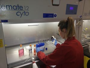
Cancer cell lines are sensitive and delicate in handling. They can only grow in the safe environment. Researchers have to protect them against bacteria, low temperatures, and too acidic/alkaline conditions. We protect cancer cells from bacteria contamination by handling them in cabinets where all plastic and media are sterile.

Handling neuroblastoma cells in the cell culture cabinet.

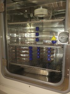
Cancer cells like to grow in conditions similar to conditions in human body. They like temperature of 36.6 – 37C. To achieve it special ‘green cell houses’ – CO2 incubators are built, which maintain the constant temperature, humidity and CO2 concentration.

We place cells in plastic dishes or containers called flasks and keep flasks in the ‘green cell houses’.

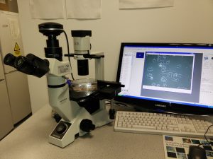
The cell growth and well being are checked regularly using microscopes. Healthy cells are to have similar shape, even distribution and grow attached to the plastic surface. Most microscopes have a camera attached to the top and linked to a computer. It helps to take picture of growing cells and record changes in cell behaviour.

Microscopic examination of drug resistant neuroblastoma cells KellyCis83. Cells look healthy and can be kept for another 2-3 days to form a more dense population.


Recommended reading

  1. Skloot, R. The Immortal Life of Henrietta Lacks 2011
  2. Thiele CJ. Neuroblastoma Cell Lines. Human Cell. 1998. 1-35 p.
  3. Murray M, Stout A. Distinctive Characteristics of the Sympathicoblastoma Cultivated in Vitro: A Method for Prompt Diagnosis. Am J Pathol. 1947;23(3):429–41.


Drug resistant neuroblastoma cells

Children with neuroblastoma undergo several cycles of intensive chemotherapy to stop disease progression with the final aim to eliminate the tumour. Chemotherapy includes carboplatin or cisplatin in various combinations with drugs such as cyclophosphamide, ifosfamide, doxorubicin, etoposide, topotecan and vincristine (1). Nevertheless, in average 1 in 5 children with stage 4 disease do not respond to therapy. Up to 50% of children that do respond experience disease recurrence with tumour resistant to multiple drugs and more aggressive behaviour that all too frequently results in death.

The development of drug resistance is the major obstacle in treatment of neuroblastoma. To tackle this problem, researchers need to study different models of disease using cell lines, 3D tumour cell models, mice models and have access to clinical samples.

The first stage in testing drugs is to understand their killing ability of cancer cells. At this stage, researchers test drugs using cell lines. Cell lines are derived from tumours which were surgically removed from children with neuroblastoma. Researchers usually take a small piece of tumour straight after surgery and bring it into the laboratory.  Here, they place this piece into special solution that has enzymes to separate cells from each other. Then the suspension of all kind of tumour cells is placed into plastic dishes or flasks in a highly nutrient media to let cells grow. Cells that can adapt to these conditions start to grow, divide and produce a new generation of cancer cells. Researchers look after their growth, inspect their shape and behaviour; and test them on the presence of tumour markers. Once identity of these cells is confirmed they become a cell line and obtain a name. These cells keep majority of characteristics of the parental tumour and represent very useful tools in cancer research.

In our lab we use such cell lines to study neuroblastoma resistance to drugs. To understand changes in neuroblastoma biology during the development of drug resistance, we created drug resistant neuroblastoma cell lines (2). We treated three neuroblastoma cell lines CHP212, SK-N-AS and Kelly with cisplatin – a common drug in anticancer therapy. SK-N-AS and Kelly cells are sensitive to this drug, while CHP212 cells responded to this drug at much higher levels that the other two. Cells were grown in media containing cisplatin for several weeks. During this period most of the cells responded to cisplatin and died. Then we let cell survivors to recover in media without drug. This cycle was repeated several times until we got a population of cell survivors that can stand doses of cisplatin that can kill 50% of parental cells.  It took us more than 6 months to generate cisplatin resistant neuroblastoma cell lines CHP212Cis100, SK-N-ASCis24 and KellyCis83.

At the next step, we studied differences between these cell lines. We first compared their behaviour and cell shapes. Two resistant cell lines KellyCis83 and CHP212Cis100 started to grow faster, but SK-N-ASCis24 – slower than their parental cell lines. Interestingly, these cells also became more resistant to other drugs such as doxorubicin, etoposide, temozolomide, irinotecan and carmustin. These results are very important as they demonstrate that one drug can activate the cell defense systems that allow to escape toxicity of other drugs. These cell lines can be used to test new drugs and find those that can overcome developed resistance.

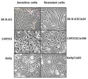
Cisplatin resistant cells also changed their appearance. Most dramatic changes occurred in SK-N-ASCis24 cells (see Figure 1).

nbl-cells

Figure 1. Microscopic images sensitive and drug resistant neuroblastoma cells (adapted from (2)) 

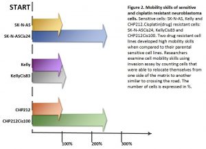
Two drug resistant cell lines SK-N-ASCis24 and CHP212Cis100 cells developed additional mobility skills – they became more invasive than their parental counterparts.


resistant-cells


Then we asked a question: what type of changes allowed cells to adapt to cytotoxic environment?  We examined changes in their genomic DNA first. We found that some genes increased their copy number, other went missing.

We identified changes in protein expression. More intriguingly, some proteins with the increased presence in the cells did not increase their presence in genomic DNA. We sorted these proteins on their role in cell processes such as migration, growth, cell cycle, etc. We found that each cisplatin resistant cell line developed a unique set of features that help them to escape cytotoxic stress (2). The similar patterns are found in clinic. Each patient responds to treatment differently.

What did we learn from this study?

  • One drug, in our study cisplatin, can activate the cell defense systems that allow to escape toxicity of other drugs.
  • The development of drug resistance gives cells new advantages and changes their behaviour and appearance, e.g. mobility skills, different cell shape, response to drugs, etc.
  • Each cisplatin resistant cell line developed a unique set of features that help them to escape cytotoxic stress.
  • These cell lines can be used to test new drugs and find those that can overcome developed resistance.

References

  1. Davidoff AM. Neuroblastoma. Semin Pediatr Surg. 2012; 21(1):2–14.
  2. Piskareva O, Harvey H, Nolan J, Conlon R, Alcock L, Buckley P, et al. The development of cisplatin resistance in neuroblastoma is accompanied by epithelial to mesenchymal transition in vitro. Cancer Lett. 2015;364(2):142–55. 


Cell to Cell Communicators

Tumour cells send different types of messages from one cell to another aka people post letters, postcards, and parcels to their families, friends, colleagues or  business. Cells can direct their messages using free moving proteins – postcards. They can wrap it in microvesicles with different cargo. Big microvesicles can take up big messages – parcels, small microvesicles or exosomes contain a limited number of texts – letters.

Tumour cells change their behaviour quickly adapting to anticancer therapies, so the messages they are sending. These messages can easily join blood stream and be read by researchers to understand how treatment is working and tumour cells are feeling.  Reading these messages from blood is more favourable as blood tests are done on the regular bases during and after the treatment.

In our lab we investigate how neuroblasts communicate with each other and the entire body through exosomes. We are interested to see what they write in their letters – exosomes. Do drug resistant and sensitive neuroblasts write different texts? What is the difference and how we can use this difference to predict child response to anticancer therapy?

In one set of experiments, we found that exosomes from drug resistant neuroblasts stimulate growth of sensitive cells. The more resistant neuroblasts send more powerful messages pushing cells to grow faster.

In the other set of experiments, we partially cracked the message showing that their texts are different. This finding explains why more resistant neuroblasts send more growth stimulating messages.

All these findings will be presented at the upcoming conference Goodbye Flat Biology: Models, Mechanisms and Microenvironment in Berlin.


schematic-exo2a

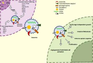
Schematic of exosome biogenesis and secretion. Cells produce exosomes through different pathways. This process is tightly regulated and controlled by numerous molecules. It can be triggered by many factors including extracellular stimuli (e.g., microbial attack, UV, drugs) and other stresses. The exosomes wrap up biologically active components such as proteins, RNA and miRNA. Exosomes can interact with recipient cells using four mechanisms: ligand/receptor interaction, protein transfer, membrane fusion or internalisation. Once exosomes entered the recipient cell, they release their content and re-programme the cell functions.


Suggested reading

Johnsen KB, Gudbergsson JM, Skov MN, Pilgaard L, Moos T, Duroux M. A comprehensive overview of exosomes as drug delivery vehicles – Endogenous nanocarriers for targeted cancer therapy. Biochim Biophys Acta – Rev Cancer. 2014;1846(1):75–87.

El Andaloussi S, Mäger I, Breakefield XO, Wood MJ a, Andaloussi S EL, Mäger I, et al. Extracellular vesicles: biology and emerging therapeutic opportunities. Nat Rev Drug Discov. 2013;12(5):347–57.

The schematic of exosomes was adapted from here

Childhood Cancer Awareness Month

September is Childhood Cancer Awareness Month!

Facts about childhood cancer

Childhood cancer is 1% of all newly diagnosed cancers globally (1,2).

It is the second most common cause of death among children under age of 19 after accidents.

Childhood cancer is an umbrella term for a great variety of malignancies which vary by site of disease origin, tissue type, race, sex, and age.

Cancer in children is not the same as cancer in adults (3–5).

The cause of childhood cancers is believed to be due to faulty genes in embryonic cells that happen before birth and develop later. In contrast to many adult’s cancers, there is no evidence that links lifestyle or environmental risk factors to the development of childhood cancer.

The most common types of childhood cancer are (1,2):

  • Leukaemia and lymphoma (blood cancers)
  • Brain and other central nervous system tumours
  • Muscle cancer (rhabdomyosarcoma)
  • Kidney cancer (Wilms tumour)
  • Neuroblastoma (tumour of the non-central nervous system)
  • Bone cancer (osteosarcoma)
  • Testicular and ovarian tumours (gonadal germ cell tumours)

In the last 40 years the survival of children with most types of cancer has radically improved owing to the advances in diagnosis, treatment, and supportive care. Now, more than 80% of children with cancer in the same age gap survive at least 5 years (1,6) when compared to 50% of children with cancer survived in 1970s-80s (7).

A revised treatment protocol was introduced in the 1970s leading to dramatic improvements in outcome for some of the most common blood cancers such as non-Hodgkin lymphoma and acute lymphoblastic leukaemia. The 5-year survival rate for non-Hodgkin lymphoma is 85% in 2003-2009. It was just less than 50% in the late 1970s. The 5-year survival rate for acute lymphoblastic leukaemia is  about 90% in 2003-2009 and just 10% – in the 1960s (1,6).Children with some types of brain cancers survive from 70% (medulloblastoma) to 85% (astrocytoma) within 5 years (2).

Unfortunately, no progress has been made in survival of children with tumours that have the worst prognosis (brain tumours, neuroblastoma and sarcomas, cancers developing in certain age groups and/or located within certain sites in the body), along with acute myeloid leukaemia (blood cancer) (1,2).  Children with a rare brain cancer – diffuse intrinsic pontine glioma survive less than 1 year from diagnosis (8). Children with soft tissue tumours have 5-year survival rates ranging from 64% (rhabdomyosarcoma) to 72% (Ewing sarcoma) (2).

For majority of children who do survive cancer, the battle is never over. Over 60% of long‐term childhood cancer survivors have a chronic illness as a consequence of the treatment; over 25% have a severe or life‐ threatening illness (9).

References:

  1. Gatta G, Botta L, Rossi S, Aareleid T, Bielska-Lasota M, Clavel J, et al. Childhood cancer survival in Europe 1999-2007: Results of EUROCARE-5-a population-based study. Lancet Oncol. 2014;15(1):35–47.
  2. Ward E, Desantis C, Robbins A, Kohler B, Jemal A. Childhood and Adolescent Cancer Statistics , 2014. Ca Cancer J Clin. 2014;64(2):83–103.
  3. Dolgin MJ, Jay SM. Childhood cancer. 1989;327–40.
  4. Miller RW, Young Jr. JL, Novakovic B. Childhood cancer. Cancer [Internet]. 1995;75(1 Suppl):395–405.
  5. Raab CP, Gartner JC. Diagnosis of Childhood Cancer. Primary Care – Clinics in Office Practice. 2009. p. 671–84.
  6. Howlader N, Noone A, Krapcho M, Garshell J, Miller D, Altekruse S, et al. SEER Cancer Statistics Review, 1975-2011 [Internet]. National Cancer Institute. 2014.
  7. Ries L a. G, Smith M a., Gurney JG, Linet M, Tamra T, Young JL, et al. Cancer incidence and survival among children and adolescents: United States SEER Program 1975-1995. NIH Pub No 99-4649. 1999;179 pp
  8. Warren KE. Diffuse intrinsic pontine glioma: poised for progress. Front Oncol [Internet]. 2012;2(December):205.
  9. Lackner H, Benesch M, Schagerl S, Kerbl R, Schwinger W, Urban C. Prospective evaluation of late effects after childhood cancer therapy with a follow-up over 9 years. Eur J Pediatr. 2000;159(10):750–8.