#JournalClubwithRonja: Reaping the benefits of PhDs past

It’s the second round of journal club blog posts, and this time around, we’ll be looking at papers published by this very lab. I’ll be focussing on the paper in which the cell line I am currently working with (KellyCis83) was developed: “The development of cisplatin resistance in neuroblastoma is accompanied by epithelial to mesenchymal transition in vitro” This research addresses a critical challenge in cancer treatment: drug resistance, of course focusing on neuroblastoma, pediatric cancer notorious for its aggressive nature and poor prognosis that this lab has been studying for years.

Neuroblastoma is typically treated with cisplatin, a potent chemotherapy drug that induces DNA damage in cancer cells, leading to their death. The issue is that over time and particularly during relapse, some neuroblastoma cells develop resistance to cisplatin, rendering the treatment ineffective. Understanding the mechanisms behind this resistance is crucial for developing new therapeutic strategies.

In this study, neuroblastoma cell lines resistant to cisplatin were created by gradually exposing the cells to increasing drug concentrations over 6 months. This approach mimics the clinical scenario where tumours are exposed to chemotherapy over an extended period, eventually leading to resistance. The resistant cell lines were then characterized to uncover the molecular changes that had occurred alongside or as part of the increased drug-resistance.

The cisplatin-resistant neuroblastoma cells exhibited significant disruptions in their cell cycle regulation, as highlighted by the most altered pathways identified by mass spectrometry. Cisplatin typically causes DNA damage that halts the cell cycle, leading to cell death. However, the researchers found upregulated pathways in resistant cells that allowed these cells to bypass this damage-induced arrest. One key finding was the identification of Vimentin upregulation in the upstream regulator analysis. Vimentin is a marker typically associated with epithelial-to-mesenchymal transition (EMT).

EMT is a process where epithelial cells acquire mesenchymal, fibroblast-like properties, including enhanced motility and apoptosis resistance. The link between EMT and cancer progression is well-established, as EMT not only facilitates metastasis but also contributes to drug resistance. In the context of neuroblastoma, the upregulation of Vimentin and dysregulation of related EMT proteins found in two of the resistant cell lines (specifically SNAI1 and TWIST1) suggests that these cells are not only evading cisplatin-induced cell cycle arrest but are also acquiring more aggressive, invasive characteristics. This links back to their findings on invasiveness, which showed greater levels in the two resistant cell lines that also had greater changes in EMT-related proteins (Figure 1).
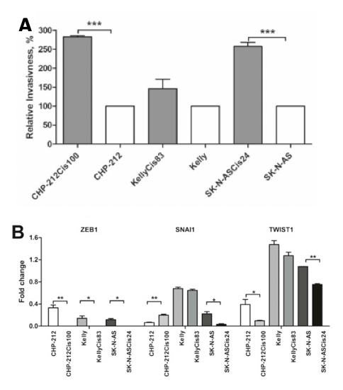
Figure 1 A Relative invasiveness of the parental cell lines compared to the cisplatin resistant daughter cell lines.  Graphed data represent mean values ± SD of three independent experiments. Asterisks indicate statistical significance obtained using a paired Student’s t-test. * p < 0.05, ** p < 0.01, ***p < 0.001, n = 3 for all experiments. B The fold change in protein expression of drug resistant cells compared to their parental counterparts was quantified by densitometric analysis of two biological repeat experiments, normalised against endogenous control ACTB. (Adapted from (Piskareva et al., 2015).

Understanding the role of EMT in cisplatin-resistance opens up new avenues for therapeutic intervention. Targeting EMT-related pathways Vimentin could potentially restore the sensitivity of these resistant neuroblastoma cells to cisplatin, by targeting the evasive mechanisms the cells developed to bypass the cell-cycle disruption. Such therapies would offer a new strategy to tackle drug-resistant relapse cases, which currently have very poor outcomes.

Overall, this study provides a valuable model for investigating drug resistance in neuroblastoma and highlights the crucial role of EMT and its associated pathways in finding ways to treat drug-resistant tumours. As we continue to explore these avenues, these models will serve us as a strong foundation facilitating the research currently taking place in our lab towards finding such combination therapies and hopefully improving outcomes for children battling this devastating cancer in the future.

Written By Ronja Struck
8th OLCHC RESEARCH & AUDIT CONFERENCE

This was our 2nd time attending the OLCHC Research & Audit Day on May 25th, 2018. The conference provides a great forum for paediatric clinicians to share and update knowledge across different specialties through talks and poster presentations. It is insightful for basic biomedical researchers like us to see other perspectives.
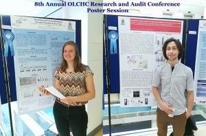
I was delighted to know that two our studies were shortlisted. It is a rewarding feeling to see your Dream Team doing very well. One was the project of the Erasmus+ student Hanne Pappaert and the other was the project of NCRC funded Postdoc John Nolan. Hanne explored our 3D tissue-engineered model of neuroblastoma using collagen-based scaffolds with distinct mechanical properties. These new scaffolds were designed and manufactured by our collaborator Dr Cian O’Leary from Pharmacy Department and Tissue Engineering and Research Group (TERG) headed by Prof Fergal O’Brien. Hanne grew 5 neuroblastoma cell lines on the 3 scaffolds: hard like a rock, soft and fluffy like a cotton wool and a jelly-like. All cells liked the jelly-like environment. This environment is similar to bone marrow – the most common site of neuroblastoma metastasis. We were excited to see the difference as it means we are one step closer to reconstruct this type of tumour spread.

John has expanded our exploration of our 3D neuroblastoma model by examining the content of exosomes – little parcels sent by cancer cells in 3D and as tumours grown in mice.  We were thrilled to see a high similarity in the exosomal content. This finding additionally proved the great applicability of our 3D model as a tool to study neuroblastoma.


Drug resistant neuroblastoma cells

Children with neuroblastoma undergo several cycles of intensive chemotherapy to stop disease progression with the final aim to eliminate the tumour. Chemotherapy includes carboplatin or cisplatin in various combinations with drugs such as cyclophosphamide, ifosfamide, doxorubicin, etoposide, topotecan and vincristine (1). Nevertheless, in average 1 in 5 children with stage 4 disease do not respond to therapy. Up to 50% of children that do respond experience disease recurrence with tumour resistant to multiple drugs and more aggressive behaviour that all too frequently results in death.

The development of drug resistance is the major obstacle in treatment of neuroblastoma. To tackle this problem, researchers need to study different models of disease using cell lines, 3D tumour cell models, mice models and have access to clinical samples.

The first stage in testing drugs is to understand their killing ability of cancer cells. At this stage, researchers test drugs using cell lines. Cell lines are derived from tumours which were surgically removed from children with neuroblastoma. Researchers usually take a small piece of tumour straight after surgery and bring it into the laboratory.  Here, they place this piece into special solution that has enzymes to separate cells from each other. Then the suspension of all kind of tumour cells is placed into plastic dishes or flasks in a highly nutrient media to let cells grow. Cells that can adapt to these conditions start to grow, divide and produce a new generation of cancer cells. Researchers look after their growth, inspect their shape and behaviour; and test them on the presence of tumour markers. Once identity of these cells is confirmed they become a cell line and obtain a name. These cells keep majority of characteristics of the parental tumour and represent very useful tools in cancer research.

In our lab we use such cell lines to study neuroblastoma resistance to drugs. To understand changes in neuroblastoma biology during the development of drug resistance, we created drug resistant neuroblastoma cell lines (2). We treated three neuroblastoma cell lines CHP212, SK-N-AS and Kelly with cisplatin – a common drug in anticancer therapy. SK-N-AS and Kelly cells are sensitive to this drug, while CHP212 cells responded to this drug at much higher levels that the other two. Cells were grown in media containing cisplatin for several weeks. During this period most of the cells responded to cisplatin and died. Then we let cell survivors to recover in media without drug. This cycle was repeated several times until we got a population of cell survivors that can stand doses of cisplatin that can kill 50% of parental cells.  It took us more than 6 months to generate cisplatin resistant neuroblastoma cell lines CHP212Cis100, SK-N-ASCis24 and KellyCis83.

At the next step, we studied differences between these cell lines. We first compared their behaviour and cell shapes. Two resistant cell lines KellyCis83 and CHP212Cis100 started to grow faster, but SK-N-ASCis24 – slower than their parental cell lines. Interestingly, these cells also became more resistant to other drugs such as doxorubicin, etoposide, temozolomide, irinotecan and carmustin. These results are very important as they demonstrate that one drug can activate the cell defense systems that allow to escape toxicity of other drugs. These cell lines can be used to test new drugs and find those that can overcome developed resistance.

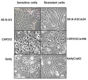
Cisplatin resistant cells also changed their appearance. Most dramatic changes occurred in SK-N-ASCis24 cells (see Figure 1).

nbl-cells

Figure 1. Microscopic images sensitive and drug resistant neuroblastoma cells (adapted from (2)) 

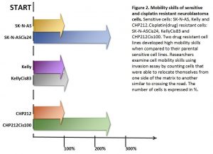
Two drug resistant cell lines SK-N-ASCis24 and CHP212Cis100 cells developed additional mobility skills – they became more invasive than their parental counterparts.


resistant-cells


Then we asked a question: what type of changes allowed cells to adapt to cytotoxic environment?  We examined changes in their genomic DNA first. We found that some genes increased their copy number, other went missing.

We identified changes in protein expression. More intriguingly, some proteins with the increased presence in the cells did not increase their presence in genomic DNA. We sorted these proteins on their role in cell processes such as migration, growth, cell cycle, etc. We found that each cisplatin resistant cell line developed a unique set of features that help them to escape cytotoxic stress (2). The similar patterns are found in clinic. Each patient responds to treatment differently.

What did we learn from this study?

  • One drug, in our study cisplatin, can activate the cell defense systems that allow to escape toxicity of other drugs.
  • The development of drug resistance gives cells new advantages and changes their behaviour and appearance, e.g. mobility skills, different cell shape, response to drugs, etc.
  • Each cisplatin resistant cell line developed a unique set of features that help them to escape cytotoxic stress.
  • These cell lines can be used to test new drugs and find those that can overcome developed resistance.

References

  1. Davidoff AM. Neuroblastoma. Semin Pediatr Surg. 2012; 21(1):2–14.
  2. Piskareva O, Harvey H, Nolan J, Conlon R, Alcock L, Buckley P, et al. The development of cisplatin resistance in neuroblastoma is accompanied by epithelial to mesenchymal transition in vitro. Cancer Lett. 2015;364(2):142–55.