Drug resistant neuroblastoma cells

Children with neuroblastoma undergo several cycles of intensive chemotherapy to stop disease progression with the final aim to eliminate the tumour. Chemotherapy includes carboplatin or cisplatin in various combinations with drugs such as cyclophosphamide, ifosfamide, doxorubicin, etoposide, topotecan and vincristine (1). Nevertheless, in average 1 in 5 children with stage 4 disease do not respond to therapy. Up to 50% of children that do respond experience disease recurrence with tumour resistant to multiple drugs and more aggressive behaviour that all too frequently results in death.

The development of drug resistance is the major obstacle in treatment of neuroblastoma. To tackle this problem, researchers need to study different models of disease using cell lines, 3D tumour cell models, mice models and have access to clinical samples.

The first stage in testing drugs is to understand their killing ability of cancer cells. At this stage, researchers test drugs using cell lines. Cell lines are derived from tumours which were surgically removed from children with neuroblastoma. Researchers usually take a small piece of tumour straight after surgery and bring it into the laboratory.  Here, they place this piece into special solution that has enzymes to separate cells from each other. Then the suspension of all kind of tumour cells is placed into plastic dishes or flasks in a highly nutrient media to let cells grow. Cells that can adapt to these conditions start to grow, divide and produce a new generation of cancer cells. Researchers look after their growth, inspect their shape and behaviour; and test them on the presence of tumour markers. Once identity of these cells is confirmed they become a cell line and obtain a name. These cells keep majority of characteristics of the parental tumour and represent very useful tools in cancer research.

In our lab we use such cell lines to study neuroblastoma resistance to drugs. To understand changes in neuroblastoma biology during the development of drug resistance, we created drug resistant neuroblastoma cell lines (2). We treated three neuroblastoma cell lines CHP212, SK-N-AS and Kelly with cisplatin – a common drug in anticancer therapy. SK-N-AS and Kelly cells are sensitive to this drug, while CHP212 cells responded to this drug at much higher levels that the other two. Cells were grown in media containing cisplatin for several weeks. During this period most of the cells responded to cisplatin and died. Then we let cell survivors to recover in media without drug. This cycle was repeated several times until we got a population of cell survivors that can stand doses of cisplatin that can kill 50% of parental cells.  It took us more than 6 months to generate cisplatin resistant neuroblastoma cell lines CHP212Cis100, SK-N-ASCis24 and KellyCis83.

At the next step, we studied differences between these cell lines. We first compared their behaviour and cell shapes. Two resistant cell lines KellyCis83 and CHP212Cis100 started to grow faster, but SK-N-ASCis24 – slower than their parental cell lines. Interestingly, these cells also became more resistant to other drugs such as doxorubicin, etoposide, temozolomide, irinotecan and carmustin. These results are very important as they demonstrate that one drug can activate the cell defense systems that allow to escape toxicity of other drugs. These cell lines can be used to test new drugs and find those that can overcome developed resistance.

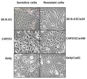
Cisplatin resistant cells also changed their appearance. Most dramatic changes occurred in SK-N-ASCis24 cells (see Figure 1).
nbl-cells

Figure 1. Microscopic images sensitive and drug resistant neuroblastoma cells (adapted from (2)) 

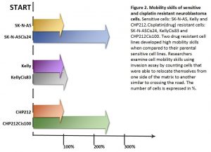
Two drug resistant cell lines SK-N-ASCis24 and CHP212Cis100 cells developed additional mobility skills – they became more invasive than their parental counterparts.
 

resistant-cells


Then we asked a question: what type of changes allowed cells to adapt to cytotoxic environment?  We examined changes in their genomic DNA first. We found that some genes increased their copy number, other went missing.

We identified changes in protein expression. More intriguingly, some proteins with the increased presence in the cells did not increase their presence in genomic DNA. We sorted these proteins on their role in cell processes such as migration, growth, cell cycle, etc. We found that each cisplatin resistant cell line developed a unique set of features that help them to escape cytotoxic stress (2). The similar patterns are found in clinic. Each patient responds to treatment differently.

What did we learn from this study?

  • One drug, in our study cisplatin, can activate the cell defense systems that allow to escape toxicity of other drugs.
  • The development of drug resistance gives cells new advantages and changes their behaviour and appearance, e.g. mobility skills, different cell shape, response to drugs, etc.
  • Each cisplatin resistant cell line developed a unique set of features that help them to escape cytotoxic stress.
  • These cell lines can be used to test new drugs and find those that can overcome developed resistance.

References

  1. Davidoff AM. Neuroblastoma. Semin Pediatr Surg. 2012; 21(1):2–14.
  2. Piskareva O, Harvey H, Nolan J, Conlon R, Alcock L, Buckley P, et al. The development of cisplatin resistance in neuroblastoma is accompanied by epithelial to mesenchymal transition in vitro. Cancer Lett. 2015;364(2):142–55. 


Cell to Cell Communicators

Tumour cells send different types of messages from one cell to another aka people post letters, postcards, and parcels to their families, friends, colleagues or  business. Cells can direct their messages using free moving proteins – postcards. They can wrap it in microvesicles with different cargo. Big microvesicles can take up big messages – parcels, small microvesicles or exosomes contain a limited number of texts – letters.

Tumour cells change their behaviour quickly adapting to anticancer therapies, so the messages they are sending. These messages can easily join blood stream and be read by researchers to understand how treatment is working and tumour cells are feeling.  Reading these messages from blood is more favourable as blood tests are done on the regular bases during and after the treatment.

In our lab we investigate how neuroblasts communicate with each other and the entire body through exosomes. We are interested to see what they write in their letters – exosomes. Do drug resistant and sensitive neuroblasts write different texts? What is the difference and how we can use this difference to predict child response to anticancer therapy?

In one set of experiments, we found that exosomes from drug resistant neuroblasts stimulate growth of sensitive cells. The more resistant neuroblasts send more powerful messages pushing cells to grow faster.

In the other set of experiments, we partially cracked the message showing that their texts are different. This finding explains why more resistant neuroblasts send more growth stimulating messages.

All these findings will be presented at the upcoming conference Goodbye Flat Biology: Models, Mechanisms and Microenvironment in Berlin.


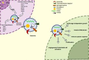
schematic-exo2a

Schematic of exosome biogenesis and secretion. Cells produce exosomes through different pathways. This process is tightly regulated and controlled by numerous molecules. It can be triggered by many factors including extracellular stimuli (e.g., microbial attack, UV, drugs) and other stresses. The exosomes wrap up biologically active components such as proteins, RNA and miRNA. Exosomes can interact with recipient cells using four mechanisms: ligand/receptor interaction, protein transfer, membrane fusion or internalisation. Once exosomes entered the recipient cell, they release their content and re-programme the cell functions.


Suggested reading

Johnsen KB, Gudbergsson JM, Skov MN, Pilgaard L, Moos T, Duroux M. A comprehensive overview of exosomes as drug delivery vehicles – Endogenous nanocarriers for targeted cancer therapy. Biochim Biophys Acta – Rev Cancer. 2014;1846(1):75–87.

El Andaloussi S, Mäger I, Breakefield XO, Wood MJ a, Andaloussi S EL, Mäger I, et al. Extracellular vesicles: biology and emerging therapeutic opportunities. Nat Rev Drug Discov. 2013;12(5):347–57.

The schematic of exosomes was adapted from here

Childhood Cancer Awareness Month

September is Childhood Cancer Awareness Month!

Facts about childhood cancer

Childhood cancer is 1% of all newly diagnosed cancers globally (1,2).

It is the second most common cause of death among children under age of 19 after accidents.

Childhood cancer is an umbrella term for a great variety of malignancies which vary by site of disease origin, tissue type, race, sex, and age.

Cancer in children is not the same as cancer in adults (3–5).

The cause of childhood cancers is believed to be due to faulty genes in embryonic cells that happen before birth and develop later. In contrast to many adult’s cancers, there is no evidence that links lifestyle or environmental risk factors to the development of childhood cancer.

The most common types of childhood cancer are (1,2):

  • Leukaemia and lymphoma (blood cancers)
  • Brain and other central nervous system tumours
  • Muscle cancer (rhabdomyosarcoma)
  • Kidney cancer (Wilms tumour)
  • Neuroblastoma (tumour of the non-central nervous system)
  • Bone cancer (osteosarcoma)
  • Testicular and ovarian tumours (gonadal germ cell tumours)

In the last 40 years the survival of children with most types of cancer has radically improved owing to the advances in diagnosis, treatment, and supportive care. Now, more than 80% of children with cancer in the same age gap survive at least 5 years (1,6) when compared to 50% of children with cancer survived in 1970s-80s (7).

A revised treatment protocol was introduced in the 1970s leading to dramatic improvements in outcome for some of the most common blood cancers such as non-Hodgkin lymphoma and acute lymphoblastic leukaemia. The 5-year survival rate for non-Hodgkin lymphoma is 85% in 2003-2009. It was just less than 50% in the late 1970s. The 5-year survival rate for acute lymphoblastic leukaemia is  about 90% in 2003-2009 and just 10% – in the 1960s (1,6).Children with some types of brain cancers survive from 70% (medulloblastoma) to 85% (astrocytoma) within 5 years (2).

Unfortunately, no progress has been made in survival of children with tumours that have the worst prognosis (brain tumours, neuroblastoma and sarcomas, cancers developing in certain age groups and/or located within certain sites in the body), along with acute myeloid leukaemia (blood cancer) (1,2).  Children with a rare brain cancer – diffuse intrinsic pontine glioma survive less than 1 year from diagnosis (8). Children with soft tissue tumours have 5-year survival rates ranging from 64% (rhabdomyosarcoma) to 72% (Ewing sarcoma) (2).

For majority of children who do survive cancer, the battle is never over. Over 60% of long‐term childhood cancer survivors have a chronic illness as a consequence of the treatment; over 25% have a severe or life‐ threatening illness (9).

References:

  1. Gatta G, Botta L, Rossi S, Aareleid T, Bielska-Lasota M, Clavel J, et al. Childhood cancer survival in Europe 1999-2007: Results of EUROCARE-5-a population-based study. Lancet Oncol. 2014;15(1):35–47.
  2. Ward E, Desantis C, Robbins A, Kohler B, Jemal A. Childhood and Adolescent Cancer Statistics , 2014. Ca Cancer J Clin. 2014;64(2):83–103.
  3. Dolgin MJ, Jay SM. Childhood cancer. 1989;327–40.
  4. Miller RW, Young Jr. JL, Novakovic B. Childhood cancer. Cancer [Internet]. 1995;75(1 Suppl):395–405.
  5. Raab CP, Gartner JC. Diagnosis of Childhood Cancer. Primary Care – Clinics in Office Practice. 2009. p. 671–84.
  6. Howlader N, Noone A, Krapcho M, Garshell J, Miller D, Altekruse S, et al. SEER Cancer Statistics Review, 1975-2011 [Internet]. National Cancer Institute. 2014.
  7. Ries L a. G, Smith M a., Gurney JG, Linet M, Tamra T, Young JL, et al. Cancer incidence and survival among children and adolescents: United States SEER Program 1975-1995. NIH Pub No 99-4649. 1999;179 pp
  8. Warren KE. Diffuse intrinsic pontine glioma: poised for progress. Front Oncol [Internet]. 2012;2(December):205.
  9. Lackner H, Benesch M, Schagerl S, Kerbl R, Schwinger W, Urban C. Prospective evaluation of late effects after childhood cancer therapy with a follow-up over 9 years. Eur J Pediatr. 2000;159(10):750–8.

A mother’s battle with neuroblastoma

A very nice piece of journalist’s story about neuroblastoma through a mother’s view. I met the mother personally at the 4th Neuroblastoma Research Symposium in Newcastle-upon-Tyne, UK in November 2015. Susan Hay and other same minded parents of children with neuroblastoma joint their efforts to raise money not only for current kids battling this nasty cancer, but more importantly for research in the cancer biology, diagnostics and new therapies which are to give a better deal for children with neuroblastoma.

https://www.theguardian.com/society/2015/nov/15/a-mothers-battle-with-neuroblastoma

Neuroblastoma summary

Neuroblastoma is a childhood cancer caused by the abnormal growth and development of non-mature nerve cells, called neuroblasts [1]. The disease commonly affects children age 5 years or younger. Approximately 50% of children have tumours that have spread at diagnosis [1]. The main challenge in treating neuroblastoma is to stop tumour spread and resistance to multiple drugs. Despite major advances in available therapies, children with drug resistant and/or recurrent neuroblastoma have a dismal outlook with 5 year survival rates of less than 20% [2-4]. Therefore, this cancer needs more research and funding as well as people awareness of these needs.


  1. Davidoff, A. M. Neuroblastoma. Semin. Pediatr. Surg. 21, 2–14 (2012).
  2. Gatta, G. et al. Childhood cancer survival in Europe 1999-2007: Results of EUROCARE-5-a population-based study. Lancet Oncol. 15, 35–47 (2014)
  3. Peinemann, F., Tushabe, D. A., van Dalen, E. C. & Berthold, F. Rapid COJEC versus standard induction therapies for high-risk neuroblastoma. The Cochrane database of systematic reviews 5, CD010774 (2015).
  4. Peinemann, F., van Dalen, E. C., Tushabe, D. A. & Berthold, F. Retinoic acid post consolidation therapy for high-risk neuroblastoma patients treated with autologous hematopoietic stem cell transplantation. Cochrane database Syst. Rev. 1, CD010685 (2015)

Welcome!

Hi readers!

This blog is about neuroblastoma biology, its research challenges, and people and media perception of this disease. I am researcher and active supporter of science communication.  I hope you will find interesting to read my blog and ask questions. Your questions would help me to cover topics which I have not heard of or may not plan to cover yet.

Welcome!