#JournalClub with Shreya: Modelling Brain Tumour Spread

This article by Krieger et al. discusses the most common form of brain cancer called glioblastoma. Due to its highly aggressive nature, research must be conducted consistently and rapidly to develop new treatments. This has proven challenging due to primary tumours being resected before further research can be done, as well as the lack of current technologies to fully explore relationships between GBM and surrounding brain tissues. This study aimed to study the aforementioned interactions in under 4 weeks, accounting for the rapid progression of the disease in real life.  

GBM cells were first derived from four patients and treated with glutamine, heparin, epidermal and fibroblast growth factors, then underwent a sequence of manipulations, such as second-generation replication lentivirus infection of GBM cells, iPSC line 409b2 inoculation in Aggrewell plates and later manipulation with invasion assays, and scRNA sequencing, which, along with the Aggrewell cells, produced neural progenitor cell spheroids for analysis. Confocal microscopy and the developed image processing algorithm allowed for visualization of these cells following fluoroscopy and depicted consistent growth of tumour cells. There was also the growth of microtubules. Any dissociated organoids were then co-cultured with GBM cells again, promoting interaction between the two. Further analysis revealed the upregulation of 45 genes, including PAX6, GJA1, GPC3, and others involved in cell regulation.  
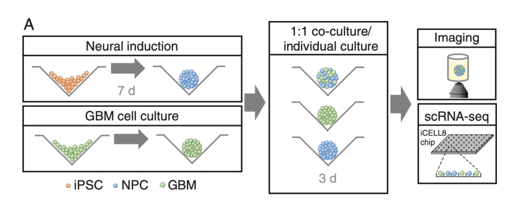
Credit to Teresa G Krieger, Stephan M Tirier, Jeongbin Park, Katharina Jechow, Tanja Eisemann, Heike Peterziel, Peter Angel, Roland Eils, Christian Conrad, Modeling glioblastoma invasion using human brain organoids and single-cell transcriptomics, Neuro-Oncology, Volume 22, Issue 8, August 2020, Pages 1138–1149

In conclusion, this novel mechanism of analysis of GBM cells using Aggrewell plates provided fruitful results, indicating intricate relationships between GBM cells and organoids, providing crucial insight for treatments by elucidating specific gene expression, heterogeneity of cells, and offering new targets based on ligand-receptor interactions. The particular relevance of this study to my work is regarding the usage of Aggrewell plates, which I am currently studying to determine how best to keep cells growing successfully within the wells. This article proves the usability and efficiency of Aggrewell and establishes its crucial role in the realm of brain cancer treatment research.  

Written by Shreya Sankar
#JournalClub with Alysia: How exosomes can promote cancer spread?

Hi, it’s Alysia, here to give you some insight on exosomes’ impact on tumor metastatic niche formation! I found an interesting paper related to my work that I want to share with you: Tumour-derived exosomes drive immunosuppressive macrophages in a pre-metastatic niche through glycolytic dominant metabolic reprogramming.

Exosomes have been a groundbreaking field of research due to their hypothesized ability to create a pre-metastatic niche through non-cancerous cell manipulation. They achieve this by relaying oncogenes and proteins from cancer cells in their molecular “cargo”. In this study, Morrissey et al. examined the role of tumor-derived exosomes (TDEs) in increasing programmed death ligand-1 (PDL-1) expression in macrophages. In pre-metastatic tumour microenvironments, there is an increase in inflammation and an immunosuppressive response, thus promoting tumorigenesis. PDL-1 is increased by TDEs manipulating the signalling of toll-like receptor 2 (TLR2) and nuclear factor kappa-light-chain-enhancer of activated B cells (NF-kB), which increases lactate production and glucose uptake in an event called the “Warburg Effect”, which is common in tumour microenvironments. TDEs taken from lung cancer samples were injected into mice, and PDL-1 expression levels of macrophages were higher than in healthy lung samples.

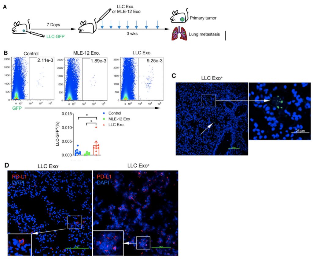
To accomplish this, Lewis lung carcinoma tumour cells (derived from mice) labelled with a green fluorescent protein (LLC-GFP+) were injected into mice with tumours; seven days later, they were injected with either non-GFP LLC exosomes or control mouse lung epithelial cell exosomes (MLE-12). The mice injected with non-GFP LLC exosomes had no metastatic difference compared to the control mice. However, the mice injected with LLC-GFP+ exosomes had an increase in micro-metastases, which was confirmed using fluorescent microscopy to observe LLC-GFP+ occurrence. With confocal microscopy, they observed an increase in PDL-1 expression in lung tissue compared to the control mice. These findings suggest that PDL-1 expression is preferentially increased in lung tumour tissue by injecting LLC-GFP+ exosomes.

Figure A. Schematic of experimental design. Figure B. Micro-metastatic burden quantified by LLC-GFP% in the lungs of s.c. GFP-LLC tumour-bearing mice treated intravenously (i.v.) with MLE-12 or LLC exosomes. Representative dot plots of GFP expression in the lungs and summarized data of LLC-GFP% are shown. Each dot represents data from one mouse. *p < 0.05, one-way ANOVA with multiple comparisons. Figure C. Confocal microscopic analysis of lung GFP+ micro-metastases from primary s.c. LLC tumor-bearing mice. Figure D. Confocal microscopic analysis of PD-L1 expression in LLC Exo- or LLC Exo+ -treated lungs. Adapted from Morrissey et al.

Researchers also examined the clinical significance of human TDEs and their manipulation of macrophages in the lymph nodes of non-small-cell lung cancer (NSCLC) patients. They found that there was increased expression of PDL-1 in the lymph nodes of these patients. To further study this, the researchers treated macrophages with either MLE-12 or LLC exosomes and then put them in a co-culture with OVA transgenic T-cells (a type of immune cell). The T-cells had a decrease in cell proliferation, which was later reversed by adding neutralizing α-PD-1 to the co-culture. This led them to think that PDL-1 had an inhibitory effect on T-cell function through macrophage stimulation. In summary, this study found that TDEs have an impact on PDL-1 expression found in tumour tissue and pre-metastatic tissue, resulting in creating an immunosuppressed environment for cancer cell growth. In my project, I’m interested in what cargo exosomes can carry from parental tumour cells to non-cancerous cells when creating a pre-metastatic niche. Knowing that there has been documented manipulation of the cellular makeup by exosomes encourages me to continue my research using neuroblastoma cells to elucidate the oncogenic markers relayed by exosomes. This research is very exciting, and I hope to shed some light on exosome impact in neuroblastoma tumour microenvironments with my own work!

Written by Alysia Scott

How things work in science: Gene editing technology

Few advancements in biomedical sciences hold as much promise for revolutionising cancer research as CRISPR-Cas9. This ground-breaking gene-editing tool has sparked a wave of innovation, offering precision and efficiency in manipulating the human genome in the fight against cancer.

Now, what is it? CRISPR is basically an acronym for a very long name Clustered Regularly Interspaced Short Palindromic Repeats Associated Protein 9 or CRISPR-Cas9 for short. It was found in simple organisms such as archaea and bacteria. Interestingly, this is a component of bacterial immune systems that can cut DNA. So, this feature was proposed for use as a gene editing tool, a kind of precise pair of molecular scissors that can cut a target DNA sequence. So, the CRISPR-Cas9 scissors allow us to precisely edit the DNA sequence of living organisms by adding in (knock-in) or removing (knockout) a gene of interest.

For cancer research, for example, the CRISPR-Cas9 scissors can be used to introduce therapeutic genes or correct mutations associated with cancer predisposition syndromes. Meanwhile, those scissors can also disrupt genes involved in treatment resistance, sensitising cancer cells to existing therapies.

Jennifer Doudna and Emmanuelle Charpentier have won the 2020 Nobel Prize in Chemistry “for the development of a method for genome editing.”. A nice accompanying piece was published in The Conversation, highlighting the history of these scissors and the politics behind it.

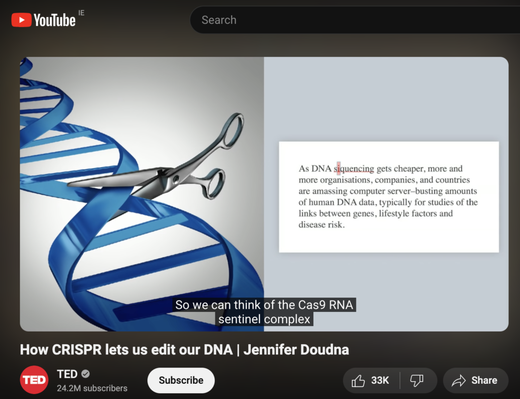
Jennifer Doudna explains this revolutionary genetic engineering tool in a TED lecture. However, she warns:

“All of us have a huge responsibility to consider carefully both the unintended consequences as well as the intended impacts of a scientific breakthrough.”

I hope you enjoyed it!

Witten by Rabia Saleem

How things work in science: targeting cell components.

How do researchers study cells? How do we get the nitty gritty?

We use many methods to tag and chase various cell components. One of my favourites is fluorescent microscopy. It allows the use of nearly all spectrum of colours from blue to purple in one go. However, we prefer to narrow it down to 2-3 colours and avoid their overlap.

How does it work? First, we use DAPI or Hoescht, which are blue fluorescent dyes used to stain DNA. This way, we tag the nucleus of the cell. Then, we tag a protein of interest. In our case, it was MYCN, a protein that acts as a transcription factor. MYCN amplification is associated with poor prognosis in neuroblastoma. As a transcription factor, it binds to genomic DNA and is located in the nucleus. We used a specific antibody that was labelled with a green fluorescent dye. Look at the image below. The green colour pattern overlaps with the blue colour. Then, we tagged the cytoskeleton, a complex of various proteins that hold the cell architecture and dynamics. We used phalloidin with red fluorescence. It is a highly selective bicyclic peptide and a popular choice for staining actin filaments.

Neuroblastoma organoids stained with DAPI, Phalloidin and anti-MYCN antibody. This work was done during the Fulbright journey to Ewald’s Lab at Johns Hopkins

Now, we can enjoy visualising cells and test different research questions. For example, how do cells respond to a drug? Or how do neuroblastoma cells spread?

Written by Olga Piskareva

How things work in science: Scaffolding

At the Cancer Bioengineering Group, we use different types of scaffolds to mimic the 3D structure of tumours outside the body. We use these scaffolds to test new therapeutics and understand the tumour microenvironment. But I bet you didn’t think we had this in common with spiders?

Spiders make their webs by producing silk from specialized glands in their abdomen. They release the silk through spinnerets located at the back of their abdomen, then use their legs to manipulate the silk strands into intricate patterns, depending on the species and purpose of the web.

The process of web building begins with a scaffold. The specialized glands that spiders use are called spinnerets, and they produce liquid silk proteins that solidify into a thread when they come into contact with air. Using their many legs, spiders can manipulate the threads by changing the speed and tension they enforce on the silk, thus controlling thickness, stickiness and strength. They first lay a framework of non-sticky threads, known as scaffolding. And layer by layer, different species of spiders will add their own artistic sticky silk design to the scaffold depending on their aim. Take the deadly redback spider for example, these guys have a utilitarian approach to web building relying on their webs mainly for shelter and capturing prey. As such, they don’t put much effort into producing irregular and messy homes. In comparison, the orb-weaving spider produces “Mona Lisa”-like designs, with complex geometric patterns and intricate designs. The differences in effort seem to come from the environments in which the webs are located, with the redbacks choosing more sheltered environments and thus not needing much strength to their webs. Whereas orb-weaving spiders are more adapted to a range of environments, from forests to grasslands to urban gardens. So, while the redback gets a lot of attention for their neurotoxic venom, they need to step up their artistic skills to match that of their orb-weaving colleagues.

The redback spider and its webs are reminiscent of an aggressive tumour, which is erratic, dangerous, and unpredictable. We want to find the “anti-venom” for such tumours so we can wipe them out for good.

Watch this amazing web-building timelapse by BBC Earth.

Written by Ellen King

How things work in Science: Tìr na nÒg

In humans, NANOG, SOX2, and OCT4 are transcription factors that maintain the undifferentiated state of embryonic stem cells (ESCs). NANOG was first discovered in 2003 by Chambers et al. and Mitsui et al. as a transcription factor in ESCs responsible for cellular self-renewal. More importantly, it enables continuous self-renewal of cancer stem cells, leading to metastasis when the regulatory genes involved do not function normally. These have been identified as cancer stem cells, with NANOG being a marker of “stemness”. In multiple cancer types, NANOG has various effects, including cellular expression of mesenchymal phenotype, cellular invasion/migration, repressed apoptosis, drug resistance, and increased angiogenesis. In pathways, NANOG either promotes or represses the expression of other genes that lead to cancer-favoured cellular behaviour. Overall, a higher expression level of NANOG is usually indicated in cases of poor prognosis.

NANOG is even more interesting due to its eponym, which comes from Tìr na nÒg. A Celtic myth of the Land of Youth, where the Tuath Dé resided in a supernatural land of paradise. This land offered beauty, health, joy, and everlasting youth to the inhabitants. As the myth goes, the Tuath Dé were gods of the land, and the god that ruled, Manannán mac Lir, was the first ancestor of humans. In various Celtic legends, humans are invited by the gods to visit Tìr na nÒg on great adventures.

However, time passes much slower in Tìr na nÒg, making it precarious for humans to return to their own world. As is the fateful tale of Oisín, who fell in love with the Tìr na nÒg goddess, Niamh. He travelled with her to Tìr na nÒg, where they lived happily in paradise. Upon a visit back to Ireland, Oisín realized that all his family had died over the years. When Oisín found a group of men who were struggling to move a giant rock, he stopped to lend them a hand while on his horse. However, the weight of the rock caused his saddle strap to snap. He fell from his horse, and when he touched the ground, he suddenly aged 300 years all at once.

Written by Alysia Scott

Sources:

Gawlik-Rzemieniewska, Natalia, and Ilona Bednarek. “The role of NANOG transcriptional factor in the development of malignant phenotype of cancer cells.” Cancer biology & therapy vol. 17,1 (2016): 1-10.

The Story of Tír Na NÓg.” Celtic Titles, 10 Feb. 2022

Women in Science: Dr. Margaret Jane Pittman

In the world of scientific research, Dr. Pittman’s name stands out. She was a bacteriologist and one of the founders of vaccines against infectious diseases such as typhoid, cholera, whooping cough and meningitis. Dr. Pittman achieved another incredible feat as the first woman to give rise to a National Institutes of Health Laboratory in the US. This accomplishment dispelled the persistent gender stereotypes that had long prevented women from pursuing careers in STEM and showcased her extraordinary credentials and experience.

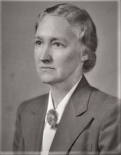
Dr. Margaret Jane Pittman was born on January 20, 1901, and grew up in Prairie Grove, Arkansas. Dr Margaret Pittman graduated magna cum laude from Hendrix in 1923. In 1925, she took her first course in bacteriology. At the time, it was propitious. Later in the nineteenth century, bacteriology became a distinct biological science. She got her Ph.D. from the University of Chicago in 1929 and started working for the National Institutes of Health in 1936. She developed, assessed, and standardised immunisation programmes against cholera, whooping cough, typhoid, and other diseases during most of her career. She continued to work at NIH as an unpaid guest. She occasionally served as a consultant for the World Health Organisation and as a guest scientist in several countries, including Spain, Scotland, Egypt, and Iran.

Her work and relentless efforts have revolutionised the world of vaccines and immunisation. Dr Pittman’s Haemophilus influenzae study has significantly influenced the medical field. Based on her research, numerous lives have been saved due to the creation of potent vaccinations, which has led to a notable drop in diseases linked to Haemophilus influenza. Pittman’s collaborations with esteemed scientists like Drs. Jonas Salk and Albert Sabin were essential in creating vaccinations against polio. They collaborated to carry out a great deal of research and clinical testing, which led to the development of the first effective polio vaccine. This enormous accomplishment was a game-changer in the global battle against polio, saving countless lives.

CREDIT: NATIONAL MUSEUM OF AMERICAN HISTORY
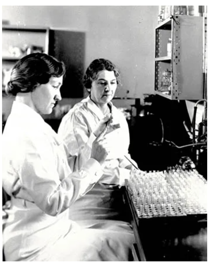
Margaret Pittman and fellow NIH physician Sadie L. Carlin are “reading” an agglutination reaction, part of the test for potency of commercially prepared anti-meningitis serum during the meningitis epidemic of 1935-1937 (1937).

As a woman in a male-dominated field, Margaret Pittman faced numerous challenges throughout her career. In an era when gender bias was pervasive, women struggled with standardisation and opportunities for advancement in scientific research. Pittman’s journey was the same, as she faced challenges that would have prevented her from moving further. The legacy of Dr. Margart Jane Pittman endures today, as her contributions continue to influence the direction of immunisation research. She stated that revolutionising problems with pertussis immunisation makes this work one of my best accomplishments that “despite the problems that have occurred with the pertussis vaccine… I consider this work one of my best accomplishments.”. Her partnerships and contributions have established a standard for upcoming generations and cleared the path for additional developments in vaccine research. By honouring her outstanding accomplishments, we can ensure that her influence on public health continues to be a driving force in the battle against infectious illnesses.

Written by Rabia Saleem

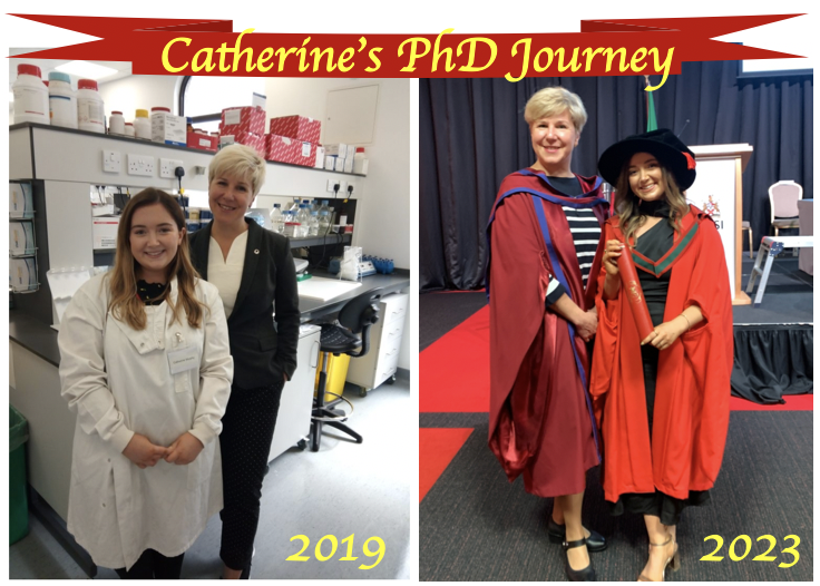
Congratulations to Dr Cat Murphy!

November 22, 2023 – Catherine was officially coined Dr Catherine Murphy. A Big Day for Catherine, her family and me.

Catherine joined our team in July 2019 to carry out a research project funded by Neuroblastoma UK. In this project, she aimed to use 3D culturing to engineer a novel experimental model and study the biology and immunology of neuroblastoma, an aggressive childhood cancer. There was the full spectrum of challenges and hard work spiced up with the uncertainty of the COVID-19 restrictions!

The PhD journey is never a straight line. It has a range of colours with 50+ shades for each. There are black alleys and hidden cul de sacs. Between July 2019 and June 2023, some days were sunny and bright, and some had scattered showers, gale winds and stormy snow, with sunshine developing elsewhere. The journey was spiced up with publications, conferences, travels, days out and fundraising events with the team.

Of note, she was behind our Twitter activities and blogging #AskCat, making our team visible! All these together have moulded into a new multi-skilled professional – Dr Catherine Murphy!

Well done to Catherine! Wish you the best of luck in your new adventure!

Women in Science: Dr. Nina Marie Tandon

Throughout history, women in science have faced the hard challenge of navigating societal biases and limited opportunities in their pursuit of scientific discovery. Many of them were unjustly denied access to education and research positions just because of their gender, as it was thought that women were not capable of rigorous scientific work.

In the present, women in science (and in many other fields) still need to face many obstacles and challenges:

  1. Gender Bias: Women in science still encounter bias and stereotypes that can hinder their progress. They may face scepticism about their abilities and qualifications, leading to a lack of recognition for their work. Gender bias can also manifest in subtle ways, affecting opportunities for advancement and funding.
  2. Unequal Pay: Gender pay gaps persist in many scientific fields, with women often earning less than their male counterparts for similar work and qualifications.
  3. Limited Representation in Leadership Roles: Women remain underrepresented in leadership roles within academia, research institutions, and industry.
  4. Work-Life Balance: Balancing a career in science with family responsibilities can be particularly challenging for women. The demands of research, long working hours, and frequent travel can conflict with traditional gender roles and family expectations.

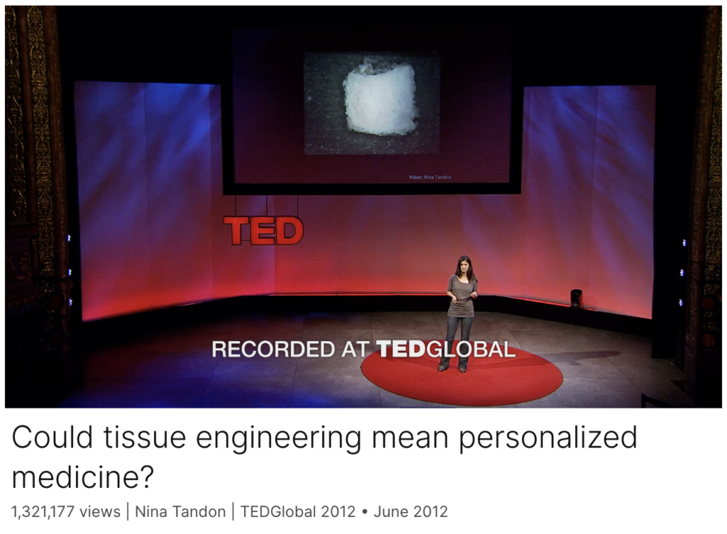
A woman and scientist who has certainly not let herself be stopped by all these adversities is Dr. Nina Marie Tandon. She has been a key player in the field of biomedical engineering, tissue engineering, and regenerative medicine. Her work focuses on developing innovative methods to grow artificial organs and tissues, using patient’s own cells to engineer tissues and organs. One of the cornerstones of Tandon’s work is the use of induced pluripotent stem cells. iPCs were developed in Japan pretty recently and are derived from skin or blood cells that have been reprogrammed back into an embryonic-like pluripotent state. This enables the development of an unlimited source of any type of human cell needed for therapeutic purposes.  

Tandon and her team employ cutting-edge bioreactors and 3D printing techniques to construct tissues: bioreactors provide a controlled environment for cells to grow and develop into specific tissue types, while 3D printing technology is used to create complex structures that mimic the architecture of natural organs.

In 2013, Tandon co-founded EpiBone, a biotech company that specializes in growing personalized bone grafts. EpiBone employs patient-specific stem cells to create skeletal structures based on individual DNA profiles, reducing the risk of rejection, streamlining surgical procedures, and potentially expediting patient recovery. They use a three-step process that begins with obtaining bone measurements and stem cells from abdominal fat via a CT scan, followed by creating a bone model in a bioreactor to stimulate growth. Finally, the patient’s own stem cells are added to the newly developed bone in the bioreactor, resulting in a fully functional replica bone ready for use.  Nina Tandon is also known for her engaging and informative TED Talks, where she discusses the future of medicine, tissue engineering, and the impact of regenerative medicine on healthcare.

I really admire Nina’s work, which I find genuinely fascinating. I believe her passion and determination come through in her TED talks, where she effectively manages to communicate complex scientific concepts and the potential of regenerative medicine to a broad audience.

Click on the image to listen to one of her TED talks. Enjoy it 🙂

Written by Federico Cottone

Women in Science: Rosalind Franklin

On a blog post series of Women in Science by a Cancer Bioengineering lab, you didn’t think you were going to get around reading about Rosalind Franklin, did you? In recent years, she finally started to receive the acknowledgement she is owed, placing her all the way up there in terms of famous scientists with Marie Curie and Albert Einstein.

As mentioned by Ellen last week, there were only ever 13 women to win the Nobel Prize in Physiology or Medicine. But in 1962, it was erroneously bestowed upon three men for the scientific breakthrough of a woman. This blog post would just as well fit into a true crime in science series.

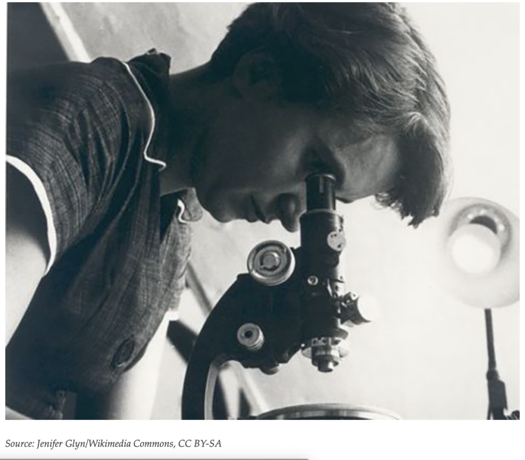
All down to misogyny and a single piece of evidence: Photograph 51, an X-ray crystallography of the structure of DNA viewed perpendicular to the DNA fibre axis, revealing the double helix structure.

You will likely all remember James Watson and Francis Crick from biology classes in school. You were probably taught that they figured out the structure of DNA.

But at the same time, they worked together in Cambridge, Franklin was working together with her PhD student Raymond Gosling at Kings College in London. Forced to work alongside Maurice Wilkins, who did not take well to her confident, goal-oriented ways, which led her to criticise her well-respected peers and dared to interrupt and correct them.

Leading to her downfall in the race with Watson and Crick was that Franklin complied with her understanding of scientific ethics, and rather than rushing to publish her findings, she sought to verify them and replicate the findings in Photograph 51.

Betrayed by her colleague Wilkins after ample tensions over the years. He passes her priceless finding to the competition, allowing Watson and Crick to model the double helix, publish a breakthrough paper and relegate her to a methods paper in the same issue of the nature journal.

Tragically, her young death at 37 from ovarian cancer prevented her from witnessing the Nobel Prize being awarded for discoveries in the molecular structure of DNA four years later. Which in 1962 was not to be awarded posthumously. Instead, her reputation for years was dominated by the more than unflattering recollections in James Watson’s biography. The book clued the public into the crimes the three men committed. All the while tarnishing Franklin’s name, portraying wildly misogynistic images and downplaying her indisputable contribution to science. Only after society as a whole changed its views on women and misogyny did perceptions of Rosalind Franklin and James Watson finally get corrected.

Today, Rosalind Franklin’s legacy stands as a symbol of tenacity, intellect, and an unyielding spirit that will inspire generations to come. It is about time that science books get rewritten to remind us that those are the virtues we should hold in high regard. Rather than the yearning for glory and a legacy that we see in the pressure to publish, the chase of impact factors never intended to rank journals and scientists but as a tool for librarians and the impossible climb through academia, forcing impossible expectations on principal investigators to take on endless students and responsibilities. Let’s take this opportunity to refocus and make sure it’s the pursuit of knowledge and answering questions that drive science forward that determine our decisions.

Written by Ronja Struck