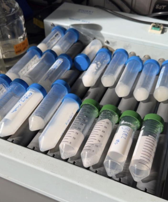
As I reflect on the work I have done leading up to my first publication since starting as a research assistant, I have thought deeply about what makes good scientific illustration, and more broadly, what makes good art. The purpose of scientific illustration is to visually synthesize research findings to aid in textual dissemination of scientific concepts. When first learning how to read a paper, I was taught to look through and try to understand each figure first. In my work process, I think about how people will look to my illustrations for clarity and understanding, as societally we translucently accept that knowledge can be and should be communicated beyond words.
There are two components that I believe make good scientific illustration. The first is understanding the language of the visual. Producing art requires integration of the subconscious into the conscious mind to communicate an idea effectively. This communication occurs in both the artist and the viewer, as the artist has to carefully consider how the viewer will both consciously and subconsciously derive meaning in an image from shape, line, form, and colour. Art that we consider ‘good’ will impact or emotionally resonate with the viewer in the way that the artist intended. Art that we consider ‘bad’ fails at communicating the meaning of its existence to the viewer.
Knowledge is what expands how we perceive beauty and what we perceive as beautiful. The second component required to create stunning visuals that effectively communicate scientific concepts and research findings to the viewers is that the artist must also have a deep understanding of the material researched. Famously, observation is the foundational principle in science and art, and curiosity is the driving factor of observation in both. Good scientific illustration must be produced by someone who intimately understands what they are drawing. Because of this, I believe that every good artist has the potential to also become a good scientist, and every good scientist has the potential to become a good artist.
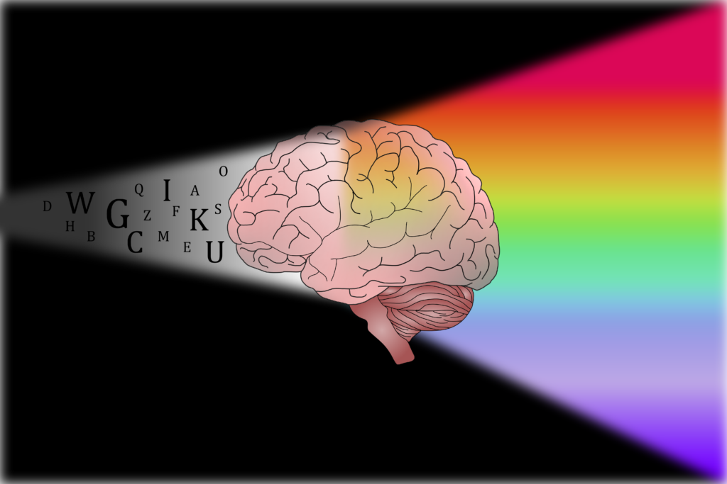
AI is something I get asked about a lot as someone who does illustration as a part of my job. We live in a world where you can ask a large-language model to generate an image summarising a text, and it is usually a bad piece of art. But why are images produced by AI often bad art? The human brain processes information through words, but also sound, image, colour, smell, touch, form, and emotion. We translate our sensory experiences and imagination into written and spoken language to communicate with each other, but we don’t always think, feel, and process in written words. All parts of living in our bodies affect our thoughts, emotions, and views of the world, and therefore, affect what we consciously and subconsciously communicate in art. Much like how dogs could not distinguish seasons solely based on how they perceive the colour of the leaves, AI fails to produce good art because it can only produce an image from the limited language of the written word.
The argument I think that a lot of people make in regards to using AI specifically in scientific illustration is that it falls into technical illustration, and therefore is inherently emotionless art intended to communicate an idea simply; sounds perfect for a robot! However, observation, a process at the very core of science, involves a synthesis of all five senses. We then primarily translate those observations into written words. Scientific illustration aims to simplify and bridge the gaps of what is lost in the written word to enhance understanding of complex concepts to a sensorially integrated mind.
As we move through this era of global uncertainty, we must befriend the liminality of it all in an attempt to reconnect with and expand our uniquely human language of art. When you make art, you are accessing a new layer of thought. I believe that when you attempt to draw your research, no matter your perceived skill level, this process helps to enhance your understanding of the material. The purpose of scientific illustration is to visually synthesise research findings to aid in the textual dissemination of scientific concepts.
Written and illustrated by Vanessa Farkas