Hot off the press! The study carried out by Thomas Frawley during his PhD has just been published in Journal of Personalized Medicine.
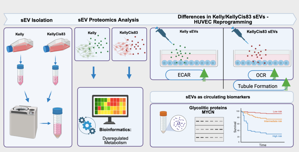
Cancer that is resistant to treatment is a big challenge because it often leads to lower survival rates. Tumour cells release small extracellular vesicles, which can influence other cells in the body by carrying various proteins. The study focused on understanding what proteins are in these particles from resistant and sensitive cancer cells and how they affect non-cancerous cells, like those involved in forming blood vessels. Our study discovered that these packages from resistant cancer cells contain special proteins involved in how cells produce and use energy. These findings suggest that these proteins could be used as markers to monitor disease progression or treatment response, using less invasive methods such as blood tests.
A schematic summary of Frawley’s study, also known as a graphical abstract. J. Pers. Med. 2025, 15(12), 584; https://doi.org/10.3390/jpm15120584 (registering DOI)
Understanding how resistant cancer cells influence their surroundings could lead to new ways of diagnosing and treating high-risk neuroblastoma. Detecting these proteins through blood tests could help personalise treatment strategies, making them more effective without the need for invasive procedures. This research opens the door to using tiny particles from blood to better understand how cancer progresses and responds to therapy.
Good afternoon, readers! Pierluca here, writing to you as one of the newest members of this incredible team. For those who haven’t met me yet, I’m a PhD student joining the RCSI family for the next three years.
My story starts in Brindisi, a charming harbor town in southeastern Italy. From there, my academic journey took me to the Netherlands. During my two research projects, I explored how high-fat diets impact liver metabolism and investigated ways to prevent metabolic reprogramming and cell death.
Now, I’m bringing that curiosity to RCSI, where my focus is shifting to something even more complex: cancer metastasis. In the lab, I’ll research how neuroblastoma invades the brain to form metastases. Using 3D bioprinting and scaffold models, I’ll grow Neuroblastoma Kelly and Kelly-cis cells to observe how they infiltrate brain-like structures and hijack the immune system.
Science is intense, so balance is key! When I’m not in the lab, you’ll find me Hiking when the sun is shining or Playing cards in a cozy pub when the rain pours. Cooking with friends is a great way to spend some relaxing time at home and when I am alone, I enjoy a good book (currently reading The Master and Margarita, highly recommend!).
Stay tuned for more about me and my research!
Written by Pierluca Cancellieri, Mac4Me PhD student
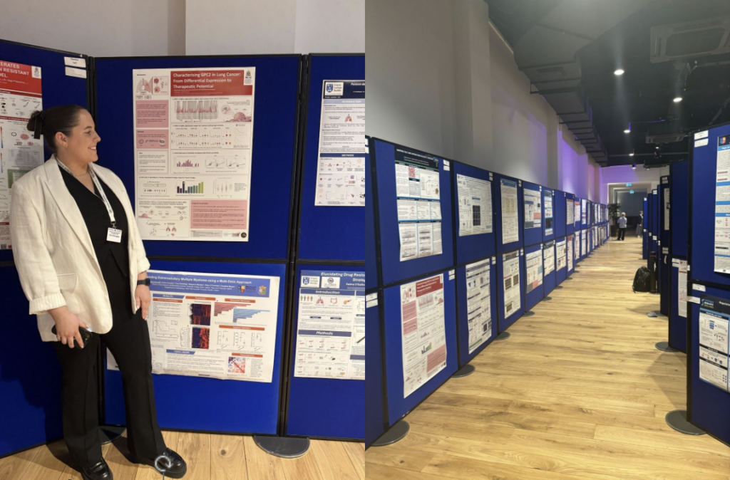
I presented a poster on my research into the regulation of GPC2 in lung adenocarcinoma and squamous cell carcinoma. The poster session was a great opportunity to share my work, receive constructive feedback, and speak with researchers working on similar topics. These conversations have given me new ideas to take back to the lab.
One of the most valuable sessions for me was the “PPI in Action” workshop, which focused on making patient and public involvement more inclusive. It introduced me to new perspectives on how researchers can engage directly with patients and the public. I left inspired to explore starting a PPI group for lung cancer at my own institution.
Another highlight was the Patrick Johnston Award session, where early-career researchers presented their work in lay terms. It was a strong reminder of the importance of clear, accessible science communication.
Beyond the conference, I enjoyed exploring Belfast—Victoria Square offered amazing views, and the architecture around City Hall was well worth the visit.
Overall, the conference was a great opportunity to connect, learn, and reflect. I’m very grateful to Breakthrough Cancer Research for supporting my attendance and look forward to applying what I learned to my research going forward.
Welcome to my first blog post of the year—and the first in two and a half years. You might be wondering what I’ve been up to during that time. Let me catch you up!
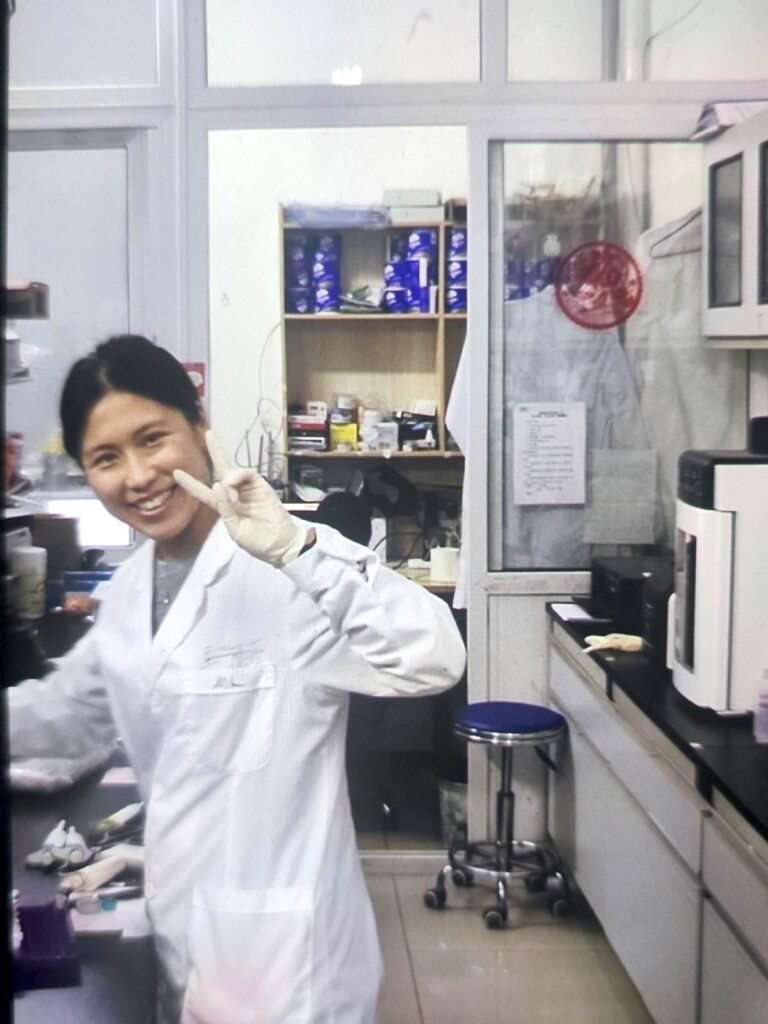
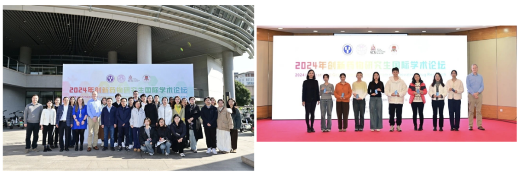
I’m currently pursuing a joint Ph.D. programme between the Royal College of Surgeons in Ireland (RCSI) and Soochow University (SU) in China. For the past two years, I’ve been based at SU. My project has offered me a unique opportunity to experience and compare research environments across two countries, each with its own strengths, workflows, and lab cultures.
So, what is it like working in a Chinese research lab?
Beyond the routine experimental tasks, one of the standout features of this lab is its comprehensive capacity for conducting animal studies—all performed in strict accordance with ethical guidelines. I’ve had the opportunity to observe and work with a wide variety of laboratory animals, including mice and rabbits. Interestingly, depending on the specific needs of a project, researchers can even select animals based on precise characteristics, such as coat colour or genetic background.
In addition to lab work, the research environment here provides frequent opportunities to attend academic conferences and participate in scholarly exchanges. These events are invaluable for sharing findings, building professional networks, and exploring future collaborations—both nationally and internationally.
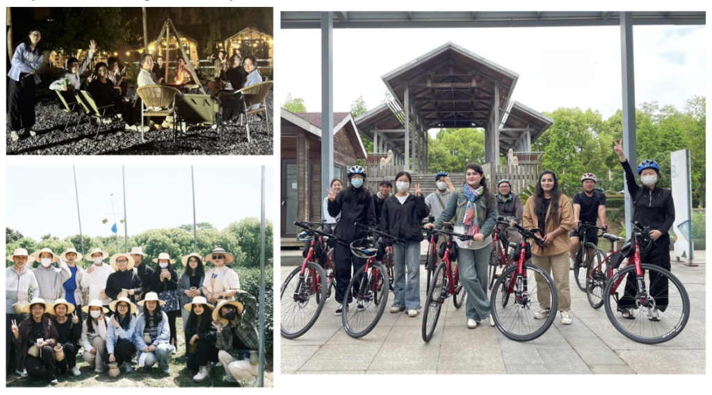
Outside the lab, work-life balance is also encouraged. My colleagues and I often take part in group outdoor activities like cycling around the nearby lake, camping, and barbecuing. These shared experiences not only bring joy to our daily lives but also help foster stronger team spirit and collaboration.
And the good news is – I have just submitted my PhD thesis! Onwards and Upwards!
I am excited to have joined the Cancer Bioengineering lab. I am passionate about studying the tumour microenvironment with the aim of understanding cellular interactions involved in tumour progression. My current work focuses on developing a 3D-printed model of prostate cancer. 3D models help us study cell-cell interactions, how cells interact with their environment, and respond to therapies.
I carried out my PhD and postdoc at University of Galway under the supervision of Dr. Aideen Ryan and Abhay Pandit, where the main focus of my project was on the optimal development of a multi-cellular 3D model of the colorectal cancer tumour microenvironment for screening colorectal cancer therapeutics. Throughout my experience, I have developed extensive skills in isolating and culturing primary cells, culturing cell lines, developing and maintaining spheroid cultures, working with a variety of hydrogels, carrying out flow cytometry, confocal microscopy and RT-qPCR. I really believe 3D models offer us great tools for understanding the tumour microenvironment and have previously developed a 3D collagen-based spheroid model of colorectal cancer, which allowed us to study the interactions between colorectal cancer cells and stromal and immune cells in the colorectal tumour microenvironment. I am really looking forward to transferring the skillset I obtained prior to working in RCSI to my project here.
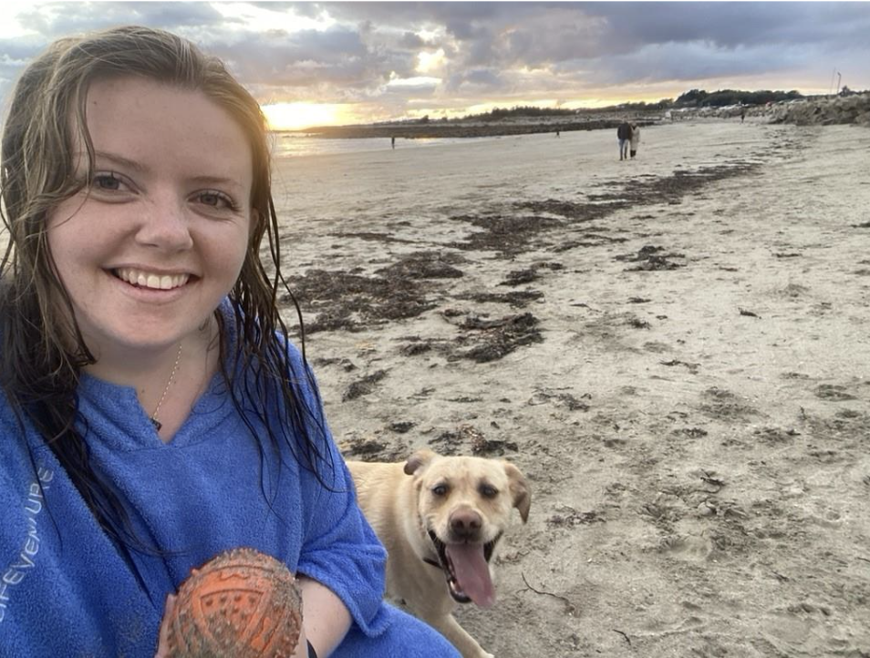
Outside of the lab, I really enjoy going for sea swims, hikes, saunas/cold plunges and going on mini road-trips around Ireland. I am looking forward to visiting all the swimming spots along the east coast of Ireland this year and am excited to contribute more to the field of cancer research.
Mac4Me goes beyond technical expertise, striving to ensure each doctoral candidate has the tools to flourish both professionally and personally. This commitment was evident in the first training, which covered clinical aspects and requirements related to the three metastatic cancer types Mac4Me is focusing on. Besides advanced scientific methodologies, including single-cell mechanics and organ-on-chip technology, the students gained insights into fundamental biological mechanisms such as tumour formation, immune evasion, and DNA repair deficiency in age-related diseases. The training also explored the ethics of cancer research and included an activity in which the communication team produced short introductory videos featuring each doctoral candidate on the website. A significant part of the training focused on Patient and Public Involvement in Research (PPI). This session, led by patient advocates from Dublin and the US, fostered an immediate connection with the doctoral candidates, emphasising the importance of collaboration and direct patient engagement at every step in the research process. A profound mutual interest in the project’s success was shared.
With nearly all doctoral candidates and principal investigators meeting in person for the very first time, the training and the meeting were marked by a palpable spirit of eagerness and enjoyment. This initial gathering fostered strong team-building among the doctoral candidates and across the various subprojects, laying a crucial foundation for their scientific and technical collaboration. The meeting proved to be a success in promoting the exchange of expertise and significantly strengthening networking opportunities, thereby setting a precedent for ongoing collaboration.
Mac4Me, Rotterdam, June 25-26, 2025
Mac4Me is a Horizon Europe MSCA (Marie Skłodowska-Curie Actions) Doctoral Network. The project is led by a core consortium of 14 partners and supported by an additional 11 associated partners. For more information about the consortium and the project, visit the Mac4Me website.
For media inquiries, please contact: mac4me@upf.edu.
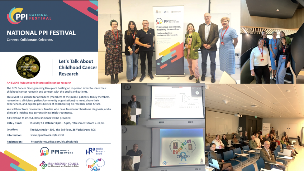
The RCSI Cancer Bioengineering Group hosted an in-person event during the National PPI Festival 2024 to share their childhood cancer research and connect with the public and patients.
We welcomed members of the public, family members of children with cancer, researchers, clinicians, and patient/community organisations on October 17th. Our past lab members and students paid a visit, too! Our group shared ongoing research on neuroblastoma biology and finding new treatments. Prof Cormac Owens from CHI brought us through the journey of clinical trials in neuroblastoma patients. We heard the heartbreaking story of the brave young man who lost his life to neuroblastoma and his parents who never gave up. This truly inspirational family founded a charity – the Conor Foley Neuroblastoma Cancer Research Foundation, to support curiosity-driven and translationally-focused research. The Foleys know very well how important it is to return happy days to kids and their families.
HDAC inhibitors are drugs that target specific enzymes involved in gene regulation. This study tested broad-spectrum HDAC inhibitors as standalone treatments and combined them with doxorubicin, a well-known chemotherapy drug.
But why on Zebrafish? The zebrafish model provides a rapid and efficient means of testing these treatments, offering valuable insights into their potential use in combating neuroblastoma. This model allows for assessing drug efficacy and helps understand the associated toxicities quickly, making it a powerful tool for developing new anti-cancer therapies.
In the study, fish larvae were implanted with fluorescently labelled, well-established neuroblastoma cell line (SK-N-BE(2)-C) and patient samples (HD-N33, NB-S-124) to grow tumours. Non-cancerous cells (VH7 fibroblasts) were utilized to verify that tumour progression in zebrafish was specific to tumour cells. The engraftment of human cells into fish larvae was confirmed by immunohistochemistry (IHC) staining on zebrafish sections injected with neuroblastoma cells (SK-N-BE). This was achieved using a STEM121 antibody that reacts specifically with a human cytoplasmic protein. The findings showed that pediatric tumour cells survive and grow in the zebrafish model at rates like those observed in human tumours.
Before testing drug efficacy in zebrafish xenografts, optimal drug concentrations and maximal tolerated doses (MTD) were determined. Toxicity tests were conducted by treating fish larvae cells for three days without tumour cell injection to identify the maximum tolerated dose that did not cause observable morbidity, changes in morphology, or severe aberrations in larval behaviour. and lethal dose (LD) for each compound. To find optimal drug concentrations, larvae with xenografted tumour cells were incubated with increasing drug doses 24 hours post-implantation to the maximally tolerated dose (MTD). The relative IC50 values were then determined based on changes in tumour mass volume.
To evaluate the treatment, SK-N-BE(2)- cells were used to test the broad-spectrum HDAC inhibitors, including panobinostat, vorinostat, and tubastatin A, both alone and combined with doxorubicin. The partial response rate (PR) was measured to see how well different drug combinations work to shrink tumours in the zebrafish model. Here’s what they found: Doxorubicin combined with panobinostat resulted in a 23% PR, Doxorubicin combined with tubastatin A showed a 31% PR, and Doxorubicin combined with vorinostat achieved the best result with a 36% PR.
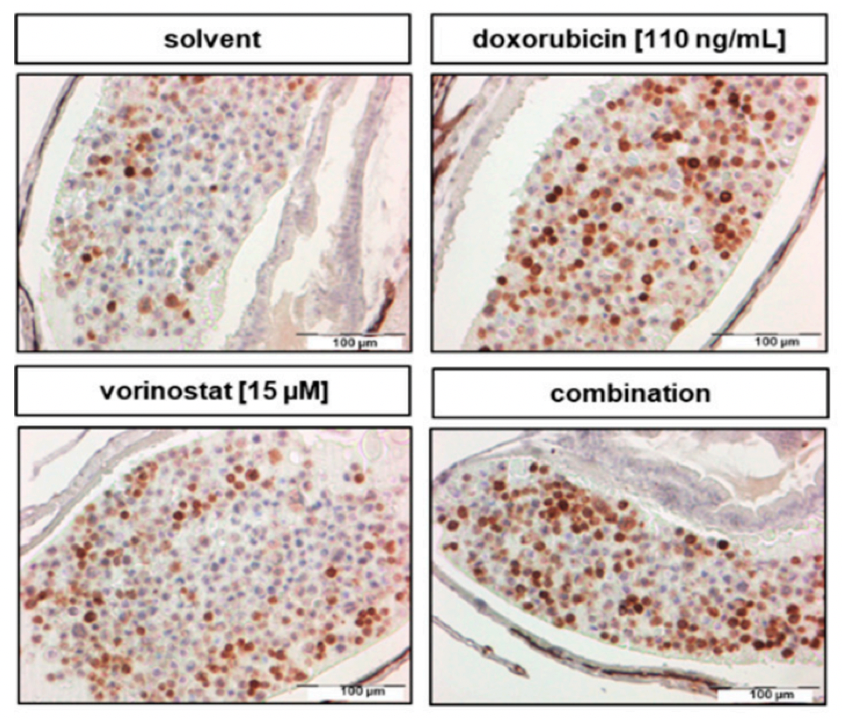
To test the effectiveness of the HDAC inhibitor treatment, they monitored the tumour growth using a confocal microscope before and 48 hours after giving the drug. The test revealed that a 48-hour treatment of SK-N-BE (2)-C zebrafish xenografts with vorinostat and doxorubicin alone, `and in combination, increased cell death. The combination of these two drugs was the most effective, causing a significant increase in cancer cell death (apoptosis) by decreasing cell proliferation, as indicated by reduced PPH3 marker and activating the number of Cleaved caspase-3 (Figure 1).
Figure 1: Treatment for 48 h with Vorinostat, doxorubicin, or a combination of both increased the amount of cleaved caspase-3 and reduced mitotic tumour cells. Adapted from Pharmaceuticals2020, 13(11), 345
In essence, this study validates the use of HDAC inhibitors in treating neuroblastoma and paves the way for broader applications of zebrafish models in cancer research. As we look to the future, these innovative models could significantly enhance our ability to develop effective cancer therapies, making strides towards better treatments and, ultimately, more effective cures.
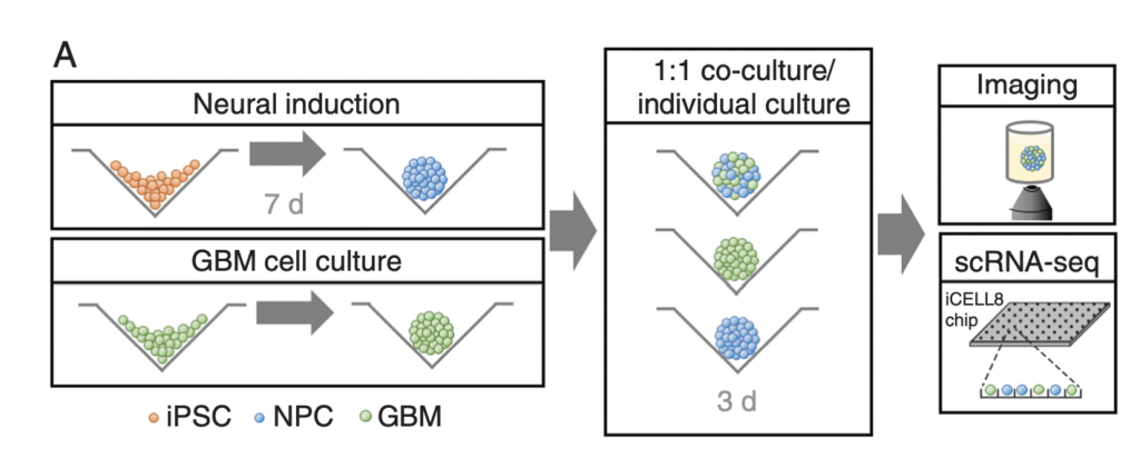
This article by Krieger et al. discusses the most common form of brain cancer called glioblastoma. Due to its highly aggressive nature, research must be conducted consistently and rapidly to develop new treatments. This has proven challenging due to primary tumours being resected before further research can be done, as well as the lack of current technologies to fully explore relationships between GBM and surrounding brain tissues. This study aimed to study the aforementioned interactions in under 4 weeks, accounting for the rapid progression of the disease in real life.
GBM cells were first derived from four patients and treated with glutamine, heparin, epidermal and fibroblast growth factors, then underwent a sequence of manipulations, such as second-generation replication lentivirus infection of GBM cells, iPSC line 409b2 inoculation in Aggrewell plates and later manipulation with invasion assays, and scRNA sequencing, which, along with the Aggrewell cells, produced neural progenitor cell spheroids for analysis. Confocal microscopy and the developed image processing algorithm allowed for visualization of these cells following fluoroscopy and depicted consistent growth of tumour cells. There was also the growth of microtubules. Any dissociated organoids were then co-cultured with GBM cells again, promoting interaction between the two. Further analysis revealed the upregulation of 45 genes, including PAX6, GJA1, GPC3, and others involved in cell regulation.
In conclusion, this novel mechanism of analysis of GBM cells using Aggrewell plates provided fruitful results, indicating intricate relationships between GBM cells and organoids, providing crucial insight for treatments by elucidating specific gene expression, heterogeneity of cells, and offering new targets based on ligand-receptor interactions. The particular relevance of this study to my work is regarding the usage of Aggrewell plates, which I am currently studying to determine how best to keep cells growing successfully within the wells. This article proves the usability and efficiency of Aggrewell and establishes its crucial role in the realm of brain cancer treatment research.
Massive congratulations on the official moulding of PhD and MSc by Research to our promising young scientists: Rabia Saleem, Dr Ciara Gallagher and Dr Ellen King! Great accomplishments!
Three different journeys, with two through the COVID-19 pandemic. The full range of ups and downs. Who said that the PhD is a straight line? It has never been. It is more like the Irish weather: some days are sunny and bright, and some have scattered showers, gale winds and stormy snow, with sunshine developing elsewhere. The journey was spiced up with publications, conferences, travels, days out and fundraising events with the team.
It is a proud moment for me as well. 🙂 Three PhD and one MSc by Research students graduated within the last 12 months.
Of note, Ellen was behind our Twitter activities in the past, making our team visible!
Wish you all the best of luck on your new adventure!