#AskLuiza: What is new in immunotherapy clinical trials for neuroblastoma?

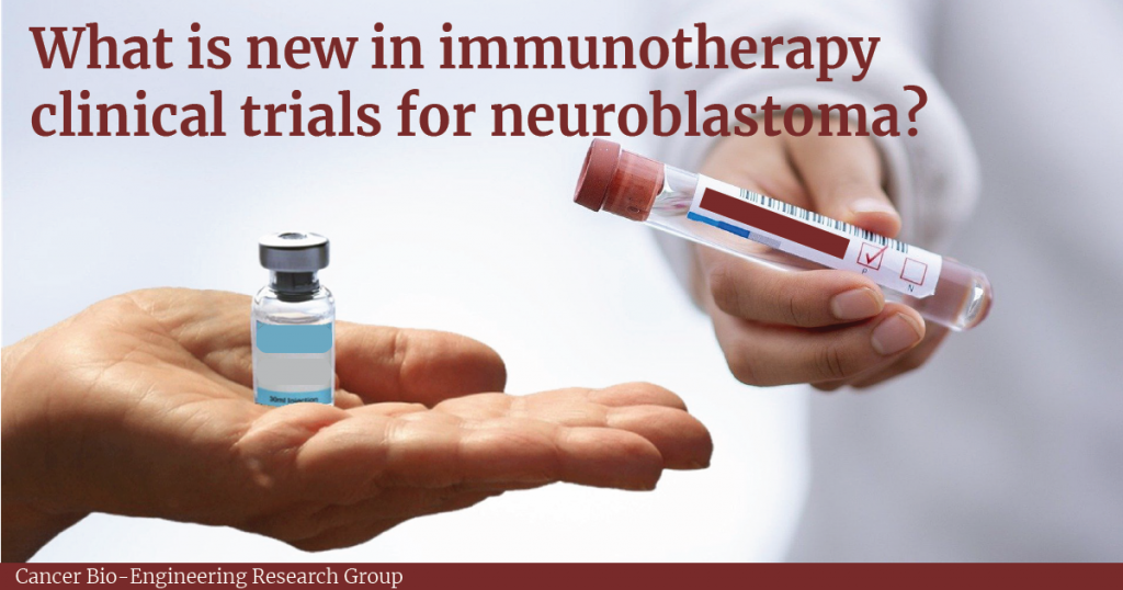
Immunotherapies are treatments that stimulate the patient’s immune system to help it to fight cancer. This type of treatment is gaining more attention in neuroblastoma due to the possibility to combine it with other therapies, potentially, generating fewer side effects.

Clinical trials are research protocols performed in patients to evaluate whether a new treatment is safe and effective. This type of research can also compare standard treatments with new treatment options as well as investigate new combinations of drugs. Clinical trials occur in phases comprising phase I (safety), phase 2 (safety and efficacy), phase 3 (safety, efficacy and comparison with standard treatments for the specific disease).

According to a search performed on November 14th, 2021, there are 594 clinical trials for neuroblastoma at clinicaltrials.gov, a clinical trial database from the United States (US). From these, 173 are recruiting or active trials and 15 are related to immunotherapies. Generally, these are initial trials evaluating treatment combinations using chemotherapy, cell transplants and immunotherapy, including antibodies and vaccines.

Trials for antibodies

The most explored target for immunotherapy in neuroblastoma is the GD2, a molecule present in the surface of neuroblastoma cells that can be used to combat the tumour. Indeed, antibodies that bind to GD2 called dinutuximab and naxitamab are approved for use in the US to treat neuroblastoma1,2.

A clinical trial in the US and Canada is recruiting patients to evaluate the combination of dinutuximab with another antibody called Magrolimab in patients with neuroblastoma that do not respond to or come back after treatment3. This is an initial trial (Phase 1), which aims to determine the best doses and side effects of this combination.

Racotumomab, an antibody that binds to N-glycolyl GM3, a molecule that is highly expressed in the surface of neuroblastoma cells, is being evaluated in high-risk neuroblastoma5. The study aims to determine the immune response generated by the drug and the related toxicity.

Trials for vaccines

A trial from Dana-Farber Cancer Institute is recruiting patients to study the GVAX Vaccine and its combination with the antibodies, nivolumab and ipilimumab, that stimulates T-cells to attack the cancer 6. The vaccine is produced with neuroblastoma cells from the patient. The study will evaluate the dose and safety of the combination treatment.

Another trial is evaluating the use of a modified neuroblastoma cell vaccine in combination with low doses of chemotherapy (Cytoxan/Cyclophosphamide)7. A vaccination scheme comprising 8 doses of vaccine and cycles of oral chemotherapy is planned and patients will be closely monitored through the vaccination period to evaluate side effects and disease status. This study is ongoing and will follow the patients for 15 years after completing the vaccination scheme.

Trials for cell therapy

A trial evaluating the use of modified T-cells (CART-T-cell) to recognise GD2- neuroblastoma cells in combination with chemotherapies (cyclophosphamide and fludarabine) and an antibody (Pembrolizumab) is ongoing8. The combination is based on previous studies that have demonstrated the longer time presence of CAR T-cell in the blood of patients after intravenous infusion of chemotherapy. Moreover, the antibody will help to stimulate the patient immune system. The trial aims to determine the highest dose possible for the combination treatment generating fewer side effects.

Another Phase I immunotherapy trial for neuroblastoma aims to compare the treatment with dinutuximab and lenalidomide (drugs that support the immune system) and Natural Killer (NK) cells from the patient9. The NK cells can kill cancer cells while the two immunotherapeutic drugs activate the NK cells. This study will determine the safest dose of cells to be used in combination with the drugs.

Conclusion

Considering some of the clinical trials in progress that uses immunotherapy to treat neuroblastoma, we can conclude that this therapy modality holds great promise to advance and potentially serve as a new treatment option to improve neuroblastoma patients’ survival and quality of life.

Written by Luiza Erthal

References

1.         Drugs Approved for Neuroblastoma – National Cancer Institute. https://www.cancer.gov/about-cancer/treatment/drugs/neuroblastoma (2011).

2.         Memorial Sloan Kettering Cancer Center. Expanded Access Use of Naxitamab/GM-CSF Immunotherapy for Consolidation of Complete Remission or Relapsed/Refractory High-Risk Neuroblastoma. https://clinicaltrials.gov/ct2/show/NCT04501757 (2021).

3.         National Cancer Institute (NCI). Phase 1 Trial of Hu5F9-G4 (Magrolimab) Combined With Dinutuximab in Children and Young Adults With Relapsed and Refractory Neuroblastoma or Relapsed Osteosarcoma. https://clinicaltrials.gov/ct2/show/NCT04751383 (2021).

4.         Memorial Sloan Kettering Cancer Center. Hu3F8/GM-CSF Immunotherapy Plus Isotretinoin for Consolidation of First Remission of Patients With High-Risk Neuroblastoma: A Phase II Study. https://clinicaltrials.gov/ct2/show/NCT03033303 (2020).

5.         Laboratorio Elea Phoenix S.A. Open-label, Multicenter, Phase II Immunotherapy Study With Racotumomab in Patients With High-risk Neuroblastoma. https://clinicaltrials.gov/ct2/show/NCT02998983 (2021).

6.         Collins, N. B. A Phase 1 Study of Combination Nivolumab and Ipilimumab With Irradiated GM-CSF Secreting Autologous Neuroblastoma Cell Vaccine (GVAX) for Relapsed or Refractory Neuroblastoma. https://clinicaltrials.gov/ct2/show/NCT04239040 (2021).

7.         Heczey, A. A Phase I/II Study Using Allogeneic Tumor Cell Vaccination With Oral Metronomic Cytoxan in Patients With High-Risk Neuroblastoma (ATOMIC). https://clinicaltrials.gov/ct2/show/study/NCT01192555 (2021).

8.         Heczey, A. Autologous Activated T-Cells Transduced With A 3rd Generation GD-2 Chimeric Antigen Receptor And iCaspase9 Safety Switch Administered To Patients With Relapsed Or Refractory Neuroblastoma (GRAIN). https://clinicaltrials.gov/ct2/show/NCT01822652 (2021).

9.         New Approaches to Neuroblastoma Therapy Consortium. A Phase I Dose Escalation Study of Autologous Expanded Natural Killer (NK) Cells for Immunotherapy of Relapsed Refractory Neuroblastoma With Dinutuximab +/- Lenalidomide. https://clinicaltrials.gov/ct2/show/NCT02573896 (2021).

#AskLuiza: How is the neuroblastoma stage determined and how does this impact treatment?

The determination of the tumour stage is an important step after a neuroblastoma diagnosis. The stage of neuroblastoma is determined depending on tumour location and if it has spread to other parts of the body. This will guide risk group assignment and treatment choice. 

The first staging system for neuroblastoma, the International Neuroblastoma Staging System (INSS), was developed in 1986 and is based on the pathological evaluation of the tumour after a removal surgery. In 2005, The International Neuroblastoma Risk Group Staging System (INRGSS) started to be used. This system is based on tumour images before any surgery. Therefore, it is based on image-defined risk factors to determine the tumour stage (see table below). It also uses clinical, pathologic, and genetic markers to determine the risk groups, which can be low-risk, intermediate-risk, or high-risk.  

Reference: Neuroblastoma – Childhood: Stages and Groups, Cancer.net

Recently, the Children’s Oncology Group (COG), a clinical trial group dedicated to paediatric cancer research revised the classification system they use to determine tumour stage for enrolment in clinical trials1. Previously, they have been defining the tumour stage based on the INNS system. Now they proposed a revised classification that takes into account the INRGSS and chromosomal alterations.  

Key clinical and biological factors used in the neuroblastoma risk classification include age at diagnosis, disease stage, tumour tissue appearance under a microscope (histology), the status of the gene MYCN that affects tumour growth, the amount of DNA in a tumour cell (called tumour cell ploidy), and alterations in the DNA.  

They analyse the outcome of almost 5,000 patients to define risk groups based on the INRGSS, using alterations in the DNA of tumour cells as a biomarker and considering current therapy modalities. In general, they found that the correlation of stages between systems is not exact. However, the differences in survival were minimal when comparing staging systems, which corroborates the use of the revised version.  

In general, the new version classifies L1 and L2 tumours as low risk, except for L1 tumours with alteration in the gene MYCN and that cannot be removed by surgery, which is high-risk. For L2 tumours, MYCN status and age can be used to evaluate prognosis. Stage M tumours can be classified as high risk or intermediate-risk depend on age, MYCN status and DNA alterations. In conclusion, low-risk groups have excellent outcomes with any or limited therapy, the intermediate-risk group have very good outcomes and high-risk groups have inferior outcomes despite therapy. 

This new version of the COG classifier will provide a uniformization of patient risk classification for clinical trials, ultimately enabling the comparison between different trials.  

Written by Luiza Erthal

Reference: 

1. Irwin, M. S. et al. Revised Neuroblastoma Risk Classification System: A Report From the Children’s Oncology Group. J. Clin. Oncol. JCO.21.00278 (2021)  

#AskLuiza: Could we use blood tests to detect neuroblastoma?

For most neuroblastoma cases a tissue biopsy, which means remove a piece of the tumour and analyse under a microscope, is performed to confirm the diagnosis, allow the risk stratification (low, intermediate or high-risk cancer) and determine treatment.  However, it is not always possible to perform a biopsy. In these cases, other tests are available such as ultrasound, x-ray and urine test1. Although these tests help the diagnostic, they are much less informative to determine the risk group and to guide treatment.

Cancer is a genetic disease and as such several genetic alterations are present in the DNA and can be useful to determine a diagnostic and prognostic of the disease. These alterations can also be useful to monitor treatment effects. But, how we can examine the DNA without using a piece of the tumour?

The answer for that relies on our blood. Cancer patients have cancer cells circulating in their blood and these cells release what we call cell-free tumour DNA. The level of cell-free tumour DNA in the blood of a healthy individual is low while this is increased in cancer patients. The detection of cell-free tumour DNA with specific alterations may help to not only detect the disease but also determine if the tumour cells are changing after treatment.

Recently, a trial for a blood test that can detect 50 different types of cancer was launched in the UK2. The test called the Galleri test look for cell-free tumour DNA in the blood using modern genetic sequencing technology. It spots the DNA that has changes common in specific cancers but not seen in healthy cells.

One of the main questions of this trial is if the test can find early stages of cancers. Although neuroblastoma is cancer that could benefit from this test, unfortunately, it is not one of the cancer types detected. However, the presence of cell-free tumour DNA was already detected in neuroblastoma patients’ blood3. Moreover, several alterations in the DNA of neuroblastoma patients have already been reported to have predictive value for disease progression and treatment monitoring. These alterations include the increase in the number of specific genes, genes breakdown or increased activity of certain genes. Their presence may indicate poor outcomes and early detection could guide to specific treatment options.

The big challenge is to have a non-invasive diagnostic method, such as blood tests, that are sensitive enough to detect the early stages of the disease. Specifically for neuroblastoma, comprehensive analysis in clinical trials regarding cell-free DNA levels and their specific changes over time would help to advance the development of liquid biopsies, such as blood tests, for this type of tumour.

Written by Luiza Erthal

References

1. Tests for neuroblastoma, Cancer Research UK. February 25, 2021

2. The Galleri multi-cancer blood test: What you need to know, Harry Jenkins, Cancer Research UK. September 13, 2021.

3. Wei, M., Ye, M., Dong, K. & Dong, R. Circulating tumor DNA in neuroblastoma. Pediatr. Blood Cancer 67, (2020).

#AskLuiza about neuroblastoma biology

#AskLuiza

We are launching a new initiative #AskLuiza to help the public and patients know more about advances and current trends in neuroblastoma.

Luiza is a research writer at the Cancer Bioengineering Research Group. She holds a PhD in Biomedical Sciences from Trinity College Dublin. You will ask a question and Luiza will look for the answer in peer-reviewed research papers that the research community trust.

Leave your question and follow our blog to read the answer soon: https://forms.gle/vrgwKoZivyUVawk9A

Models to study neuroblastoma in the laboratory

Finding suitable research models to study disease is a big challenge for researchers around the world. In cancer research, it is essential to work with models that can recapitulate tumour characteristics as much as possible. This is important to test chemotherapeutic drugs, understand tumour behaviour and have higher chances of translating the finds from the laboratory to clinical practice.  

Multiple factors influence tumour behaviour and disease progression. The most important is the tumour microenvironment, which comprises different cells and molecules that surround the tumour and the extracellular matrix, a network of molecules that provides support to the cells in the body.  

Most cell studies in a laboratory are based on 2D cell culture models in which the cells grow in a monolayer. Although this approach has a low cost and it is easy to use, it lacks the complexity observed in the clinical scenario. It is true that no model can recapitulate all the complexity found in the body. However, scientists were able to develop interesting approaches to study different tumour characteristics with relatively good approximation1.  

Specifically for neuroblastoma, the most common solid tumour that affects children, scientists developed 3D models in which neuroblastoma cells grow interacting with the surrounding environment and with each other in a vial. Examples of 3D models include cells grown in hydrogels or scaffolds and multicellular tumour spheroids (see image below). Spheroids are formed through the self-adhesion of tumour cells growing in the form of very small balls. They can be maintained in the laboratory on their own or supported by scaffold-based platforms (jelly-like or porous materials). Scaffolds essentially support the cell resembling the extracellular matrix and surrounding tissue in the body. 

In the Cancer Bioengineering Research Group, we work with neuroblastoma models such as organoids, a more complex type of spheroid, to understand neuroblastoma migration and invasion2. Moreover, we recently shared with the research community a protocol at jove.com describing the development of a 3D neuroblastoma model using collagen-based scaffolds3.  

Time-lapse video of neuroblastoma organoids’ growth. Accompanying experimental data published in Gavin et al., Cancers 2021. Source: the Cancer Bioengineering Research Group 

These models have the potential to advance drug tests performed in the laboratory providing better clinical translation, ultimately contributing to improving the quality of life and survival of children diagnosed with neuroblastoma.  

The work with 3D models at the Cancer Bioengineering Research Group is supported by the Irish Research Council, the Conor Foley Neuroblastoma Cancer Research Foundation, Neuroblastoma UK and National Children’s Research Centre. 

Written by Luiza Erthal

References 

1. Nolan, J. C. et al. Preclinical models for neuroblastoma: Advances and challenges. Cancer Lett. 474, 53–62 (2020). 

2. Gavin, C. et al. Neuroblastoma Invasion Strategies Are Regulated by the Extracellular Matrix. Cancers 13, 736 (2021). 

3. Gallagher, C., Murphy, C., O’Brien, F. J. & Piskareva, O. Three-dimensional In Vitro Biomimetic Model of Neuroblastoma using Collagen-based Scaffolds. J. Vis. Exp. 62627 (2021) doi:10.3791/62627.