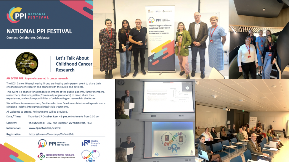
The RCSI Cancer Bioengineering Group hosted an in-person event during the National PPI Festival 2024 to share their childhood cancer research and connect with the public and patients.
We welcomed members of the public, family members of children with cancer, researchers, clinicians, and patient/community organisations on October 17th. Our past lab members and students paid a visit, too! Our group shared ongoing research on neuroblastoma biology and finding new treatments. Prof Cormac Owens from CHI brought us through the journey of clinical trials in neuroblastoma patients. We heard the heartbreaking story of the brave young man who lost his life to neuroblastoma and his parents who never gave up. This truly inspirational family founded a charity – the Conor Foley Neuroblastoma Cancer Research Foundation, to support curiosity-driven and translationally-focused research. The Foleys know very well how important it is to return happy days to kids and their families.
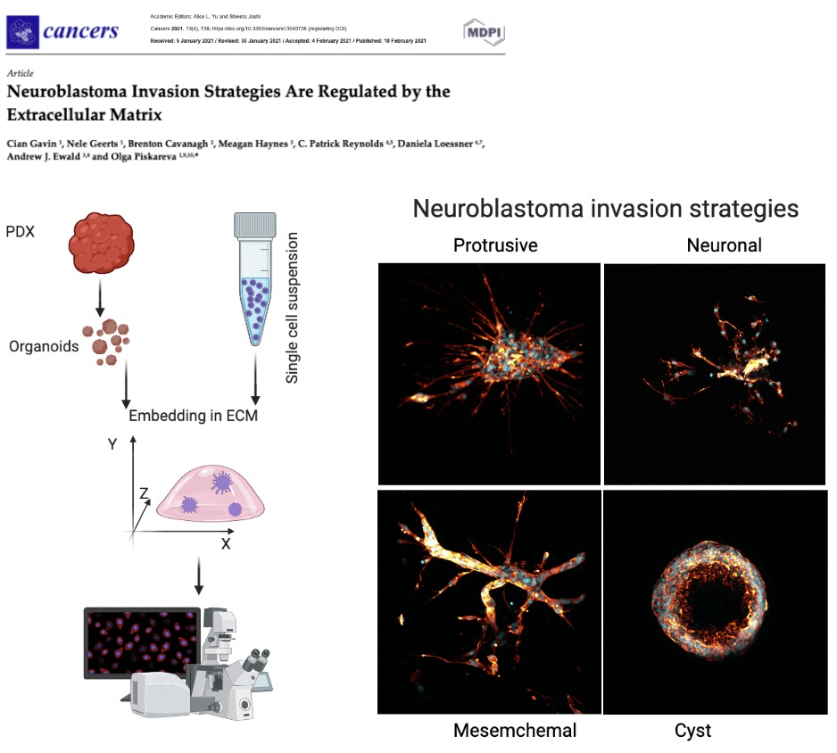
As a new PhD student, I’m incredibly excited to dive into cancer research, and what better way to kick off this journey than by exploring 3D models to study neuroblastoma metastasis? Neuroblastoma is one of the most common childhood cancers, and about 50% of patients have metastatic disease at diagnosis. Understanding how these cells spread is key to developing better therapies, which is why this recent study by Gavin et al. (2021) caught my eye.
So, what did the researchers do? They used something called patient-derived xenografts (PDX) and cell lines to grow organoids (tiny mini-tumors) in a 3D extracellular matrix (ECM). This ECM mimics the environment these cells would encounter in the body, which is super important because cells behave very differently in 3D than in the typical 2D Petri dishes. It’s like giving the cells an entire landscape to explore rather than just a flat road—suddenly, they have mountains to climb and valleys to cross, allowing them to behave much more like they would inside the body!
One of the coolest things about this study is how the neuroblastoma cells developed various invasion strategies based on their environment. Some stayed in tightly knit groups, while others decided to go full-on lone wolf, sending out long, thin projections to explore the surrounding matrix. These cells are smart-adapting to different ECM compositions like Matrigel (which is rich in laminin and collagen), made them change their behaviour entirely. It’s like they’re navigating an obstacle course, with each new challenge requiring a different tactic!
Let’s Talk Actin Filaments!
Now, this is where it gets super cool (and nerdy in the best way!). The images captured by confocal microscopy are stunning. They show actin filaments—the internal skeleton of the cells—as they help the cancer cells move and invade new areas. The actin filaments form these amazing, intricate networks that shape the cells and allow them to stretch and invade. It’s almost like watching tiny construction workers build bridges and tunnels as they move forward. Check out this confocal image showing the red filaments—how awesome is that?!