Hi everyone! I’m Chunyu

Hi everyone! I’m Chunyu, and I’ve recently started my PhD journey in the field of bioengineering and neurobiology. My academic background includes an MRes in Biomedical Research from Imperial College London, where I developed a deep interest in microfluidic technologies and their applications in disease modelling. 

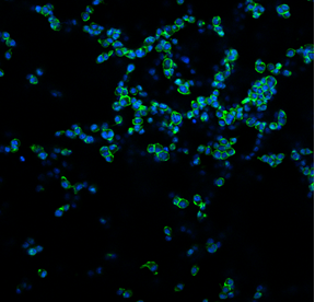
Currently, my PhD project focuses on identifying the function of macrophages—the body’s frontline immune cells—when they first interact with neuroblastoma (NB) cells using a brain and liver organ-on-a-chip (OoC) model. By recreating these organ environments on a chip, I aim to explore how macrophages respond to NB invasion and how this early interaction might shape the progression of the disease. This research could open new doors for early intervention and treatment strategies in childhood cancers like neuroblastoma. 

When I’m not in the lab, you’ll probably find me outside—going on hikes, enjoying a good swim, or finding a tasty Hotpot restaurant. I love blending my curiosity for science with a love for the outdoors, and I’m excited to share updates from both worlds as I go through this PhD journey. 

Thanks for stopping by, and stay tuned for more science and a few outdoor adventures along the way! 

Written by Chunyu Yan, Mac4Me DC

Colouring cells in research

Sometimes, the most fascinating parts of science are invisible to the naked eye—like in these images captured with a confocal microscope! 

What you’re seeing here are DC 2.4 cells, a mouse dendritic cell line. These immune cells are key players in recognising foreign substances (like bacteria, viruses, or even cancer cells) and activating the body’s immune response. 

In this experiment, we cultured the DC 2.4 cells on a sponge-like material composed of collagen and glycosaminoglycans (GAG), two natural components commonly found in body tissues. This material is called a scaffold, and it provides cells with a 3D surface to grow on, more closely mimicking their natural environment within the body. 

To make the cells visible under the microscope, we used two fluorescent stains: 

  • DAPI (blue), which marks the nucleus—the control centre of the cell, 
  • Phalloidin (green), which highlights the actin filaments that give the cell shape and structure. 

We’re testing how well these immune cells survive, attach, and spread on the collagen-GAG scaffold over time. By utilising a 3D environment, we can gain a deeper understanding of how cells behave in more realistic conditions. This is especially important for research into cancer immunotherapy and vaccine development. 

This image tells us that the DC 2.4 cells can successfully grow and interact with the scaffold! 

Written by Federica Cottone