

Sometimes, the most fascinating parts of science are invisible to the naked eye—like in these images captured with a confocal microscope!
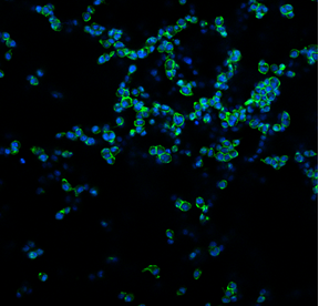
What you’re seeing here are DC 2.4 cells, a mouse dendritic cell line. These immune cells are key players in recognising foreign substances (like bacteria, viruses, or even cancer cells) and activating the body’s immune response.
In this experiment, we cultured the DC 2.4 cells on a sponge-like material composed of collagen and glycosaminoglycans (GAG), two natural components commonly found in body tissues. This material is called a scaffold, and it provides cells with a 3D surface to grow on, more closely mimicking their natural environment within the body.
To make the cells visible under the microscope, we used two fluorescent stains:
- DAPI (blue), which marks the nucleus—the control centre of the cell,
- Phalloidin (green), which highlights the actin filaments that give the cell shape and structure.
We’re testing how well these immune cells survive, attach, and spread on the collagen-GAG scaffold over time. By utilising a 3D environment, we can gain a deeper understanding of how cells behave in more realistic conditions. This is especially important for research into cancer immunotherapy and vaccine development.
This image tells us that the DC 2.4 cells can successfully grow and interact with the scaffold!
Written by Federica Cottone