Models to study neuroblastoma in the laboratory

Finding suitable research models to study disease is a big challenge for researchers around the world. In cancer research, it is essential to work with models that can recapitulate tumour characteristics as much as possible. This is important to test chemotherapeutic drugs, understand tumour behaviour and have higher chances of translating the finds from the laboratory to clinical practice.  

Multiple factors influence tumour behaviour and disease progression. The most important is the tumour microenvironment, which comprises different cells and molecules that surround the tumour and the extracellular matrix, a network of molecules that provides support to the cells in the body.  

Most cell studies in a laboratory are based on 2D cell culture models in which the cells grow in a monolayer. Although this approach has a low cost and it is easy to use, it lacks the complexity observed in the clinical scenario. It is true that no model can recapitulate all the complexity found in the body. However, scientists were able to develop interesting approaches to study different tumour characteristics with relatively good approximation1.  

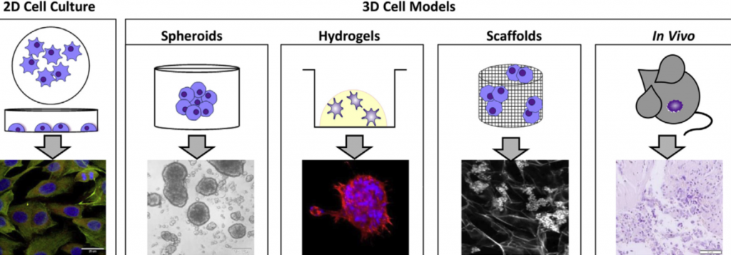
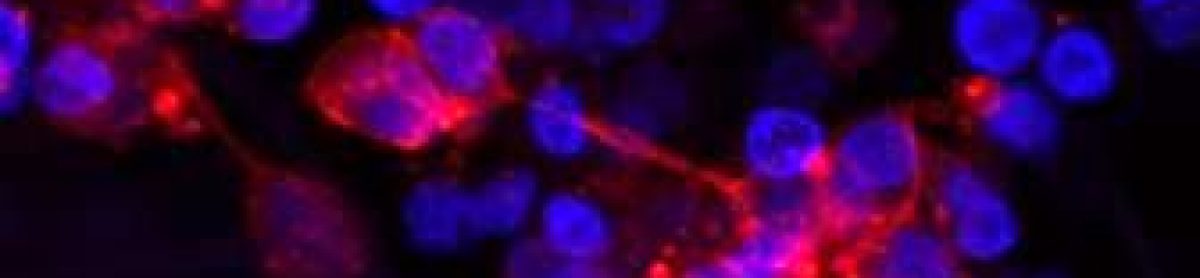
Specifically for neuroblastoma, the most common solid tumour that affects children, scientists developed 3D models in which neuroblastoma cells grow interacting with the surrounding environment and with each other in a vial. Examples of 3D models include cells grown in hydrogels or scaffolds and multicellular tumour spheroids (see image below). Spheroids are formed through the self-adhesion of tumour cells growing in the form of very small balls. They can be maintained in the laboratory on their own or supported by scaffold-based platforms (jelly-like or porous materials). Scaffolds essentially support the cell resembling the extracellular matrix and surrounding tissue in the body. 

In the Cancer Bioengineering Research Group, we work with neuroblastoma models such as organoids, a more complex type of spheroid, to understand neuroblastoma migration and invasion2. Moreover, we recently shared with the research community a protocol at jove.com describing the development of a 3D neuroblastoma model using collagen-based scaffolds3.  

Time-lapse video of neuroblastoma organoids’ growth. Accompanying experimental data published in Gavin et al., Cancers 2021. Source: the Cancer Bioengineering Research Group 

These models have the potential to advance drug tests performed in the laboratory providing better clinical translation, ultimately contributing to improving the quality of life and survival of children diagnosed with neuroblastoma.  

The work with 3D models at the Cancer Bioengineering Research Group is supported by the Irish Research Council, the Conor Foley Neuroblastoma Cancer Research Foundation, Neuroblastoma UK and National Children’s Research Centre. 

Written by Luiza Erthal

References 

1. Nolan, J. C. et al. Preclinical models for neuroblastoma: Advances and challenges. Cancer Lett. 474, 53–62 (2020). 

2. Gavin, C. et al. Neuroblastoma Invasion Strategies Are Regulated by the Extracellular Matrix. Cancers 13, 736 (2021). 

3. Gallagher, C., Murphy, C., O’Brien, F. J. & Piskareva, O. Three-dimensional In Vitro Biomimetic Model of Neuroblastoma using Collagen-based Scaffolds. J. Vis. Exp. 62627 (2021) doi:10.3791/62627. 

I’m Ronja

I’m Ronja, I’m from Germany, but have spent my entire adult life in the English-speaking parts of this world. Right after school, I interned with a PhD student working on cystic fibrosis for a couple of months. Having the chance to culture airway epithelial cells myself made me certain I was on the right track with biomedical sciences. So, I studied Biomedical Sciences (Anatomy) in Aberdeen. The best part of that degree was my introduction to dissections. I enjoyed them so much that I even considered becoming a full-time prosector. But that does not count as essential work, so I found a remote master’s degree in Health Research instead. Studying remotely gave me the fabulous opportunity to structure my own time. I could go and explore Scotland during the day and work in the evenings. But after 5 years of studying, I was finally ready to start a PhD and was ever so delighted when I heard I could weave in some dissections at RCSI. Now, I’m looking forward to discovering what Dublin has to offer and to getting stuck in my research project!

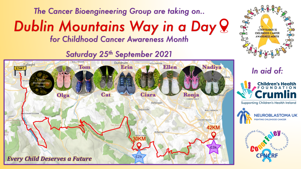
While I didn’t know much about the particulars of my PhD before starting, I had an idea about the project from the application and I knew accommodation was sorted out for me, but I had never seen the place or my future flatmates. The one thing that I was made aware of far in advance of moving to Dublin was that September was Child Cancer Awareness Month, for which the team was going to do a charity event. Based on past years I was expecting it to be a 10km run, which was pretty daunting to me. So, I prepared. I started running and cycling over the summer until 10km weren’t an issue anymore. But in the first lab meeting plans shifted. We were going to do the Dublin Mountains Way in a day. The 10km were tripled and depending on donations maybe even quadrupled. Quite a different challenge! But I believe my summer prepared me well for that too. Alongside running I started cycling a little as well. And because there was a free bike in Aberdeen for me, I cycled it down to Stirling. Let’s hope that the endurance needed to cycle 200km translate to hiking 30-42km!

Ronja Struck

Dublin Mountain Way in A Day, September 25th 2021

Here are our plans. This year we have upped the challenge, taking on the Dublin Mountain’s Way in a Day ⛰ We will hike through the Dublin Mountains from Tallaght to Glencullen, and maybe even all the way to Shankill on September 25th! Our challenge is not only to do #DMW in a Day & support three wonderful charities CMRF Crumlin/National Children’s Research Centre, Neuroblastoma UK and the Conor Foley Neuroblastoma Cancer Research Foundation but also beat our past fundraising records! If we raise 2K+, we’ll do 30km in a day. If 3K+ then 42km! Can u challenge us?  All funds raised will go to the 3 selected charities. Every donation big or small is hugely appreciated!

Please support us by donating to our Gofundme

https://gofund.me/ec59f131

One Day of the Life as a Researcher: PhD student

You may wonder whether I re-submit the same. the third time? Actually, our team has 3 ongoing PhD students and one starting from October. So, here we are. Three identical titles so far but different journeys. Today, it is Tom’s turn to tell his story.

Three years I ago I decided to try my hand at some cancer research and quit my job as a medical scientist in a diagnostic lab. I am now in my third year of a PhD and I am certain I made the right choice. It was a challenging transition from working in an environment with a lot of automation and standard operating procedures to one where you have to figure out everything for yourself! However, I think that that learning experience has allowed me to adjust quite well to all of the COVID-19 related upheaval. 

Pre-pandemic you could saunter between your office and the lab as often as you pleased, you had a choice of at least four different places to go for coffee on campus and you could squeeze into a packed lift to avoid the stairs to the lab. Now a day in the lab is quite different. We have to book lab space online, social distance from our colleagues, frequent hand washing and wear a mask at all times.

Tom Frawley

These days I plan all my lab work and book lab bench space the week before. On a typical day I split my time between the lab and working from home. I am quite fortunate that my commute is only a 6 minute walk through Stephens Green, which is only 5 minutes longer than the walk from the lab to my old office. 

Working through a pandemic is certainly challenging however I do appreciate my time in the lab much more now and I feel like I am much more productive when access to the lab is limited. 

Thomas Frawley, the IRC-NCRC funded PhD student.

One Day of the Life as a Researcher: summer undergraduate student

This summer I worked under Dr Piskareva supervision in the remote research program. My original plans for a lab experience were put down by COVID. My ultimate goal was to write the review article on the potential uses of retinoic acid in neuroblastoma research.

Nadiya Bayeva

Before starting the project, I didn’t have any specific expectations. While I always had fun picking the primary articles apart and summarising the gathered data during my undergrad and med school, I didn’t believe that this experience would be the special one. And I kept thinking so as I was collecting the information from the numerous data on cell cultures. And as I was looking at the mice models studies. And clinical trials.

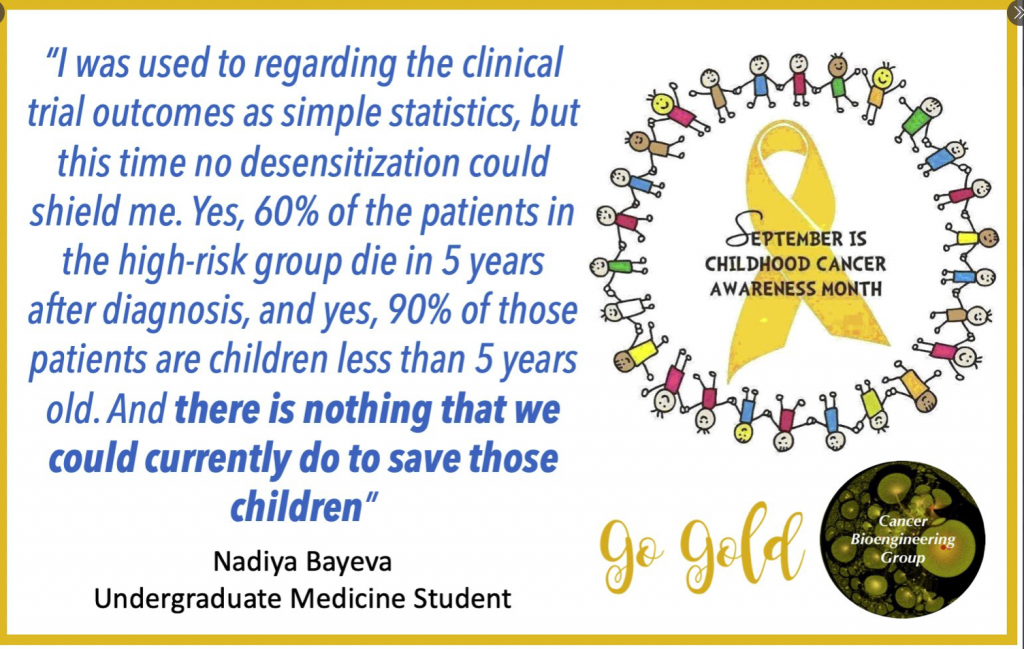
Then I started to write my introduction, and so researched the information on the neuroblastoma prognosis, contemporary treatment protocol and outcomes. And suddenly I saw my project in the new light. I was used to regarding the clinical trials outcomes as simple statistics, but this time no desensitization could shield me. Yes, 60% of the patients in the high-risk group die in 5 years after diagnosis, and yes, 90% of those patients are children less than 5 years old. And there is nothing that we could currently do to save those children.

On the other hand, this realisation brought meaning to my work. This time, I was not doing a PubMed search to get a good mark or CV reference. Instead, I was looking for the possible treatment of the disease. My review on the current knowledge about retinoic acid will let the other primary researchers target the most promising drug for future experiments and, eventually, create a novel and effective therapy to help those children.

And isn’t it what we are all striving for in medical research?

International Childhood Cancer Awareness Day – February 15th 2019

International Childhood Cancer Day (ICCD) was founded in 2002 by Childhood Cancer International (CCI). Each year on February 15th we unite together to recognise childhood cancer as a national and global child health priority & to raise support, funding and awareness of this devastating desiease.

This year we team up with Amorino to run Hot Chocolate Morning.  Please come along! All proceeds go to CMRF/NCRC and CFNCRF.

If you can’t join us, you can simply follow the link and donate ‘a cup of coffee/hot chocolate’ to CMRF Crumlin, the Conor Foley Neuroblastoma Research Foundation & Childhood Cancer Foundation


Scientific part of my journey

Reading my posts, it looks like I am more enjoying the cultural part and almost forgot the main reason I crossed the Atlantic with the Fulbright wings.

The first month in the lab was more a warming up. Where is my desk? Where is the cell culture rooms? How do they run it? How different is it? So, many microscopes – am I capable of imaging? And so on and so forth…

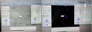
My typical day starts at 8-8.30 am and finishes once all is done. It may be 6pm or 10pm. Once the experiment is set up, I have to monitor cells every 24 hours for 5-7 days with no weekends or days off. The monitoring includes imaging. Lots of imaging. Every condition has 20-30 single cells to follow up. Each cell gets its own GPS tag manually to be able to image exactly the same cell as it grows and becomes a group of hundreds by multiplication. For example, I am running 8 different cell lines in 3 experimental conditions. So, 20-30 cells per all 24 combinations give us 480-720 individual cells to follow up. The imaging takes ~5 hours every day. After 5 days, I will have 2400 – 3600 pics of cells to analyse. It will be fun! I may need lots of Guinness to fly through that numbers.

Tagging cells. The left arrow points to a group of neuroblastoma cells. The arrow in the middle point to the same cells, but this image allows you to see the actual number of the cells. This group has 8 cells. The right arrow points to individual GPS tags for each cell

At the next step, I will select some of the conditions for video recording to trace cell fate from a single neuroblastoma cell to a metastatic niche consisting of hundreds of them. This video will show me how it all happens minute after minute.

Is not it exciting? I am thrilled!


Research Summer School 2018

Another year, another Research Summer School students. We are hosting 4 students (Jessica, Dawn, Dola, and Jeff) this year. Some of them will be medical doctors, another will do research after the graduation. For them, the 8-weeks lab placement is a window into the reality of the everyday science. How cancer cells look? How do they grow? Where do we store them? How do we know that we have identified a new drug or a new target to study further? Do researchers have a sense of humour? Do they like donuts?

Why do they wear these astronaut helmets?

We have already said Good Bye to Jessica. Dola and Dawn’s projects are coming to an end this week, while Jeff is staying till the end of August.

How current IT advances help in research?

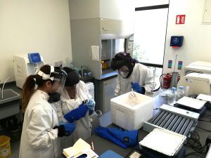
Here is the perfect example of the teamwork troubleshooting protein extractions. My Dream Team 2018 in action. The current information and communication technologies allow to stay connected and respond quickly.

Five minutes later in the lab: troubleshooting is the exchange of experiences!


A new 3D strategy to study neuroblastoma

Our body has 3 dimensions: height, width and depth. Every single part of our body grows in the same 3 dimensions. This is true for cancer cells. Researchers use different ways to study cancer cells behaviour, how they grow and spread. We grow cells in the flasks, where they change their structure and shape and become flat losing one dimension. This is a very popular approach. We also grow cells in mice, where cells keep their 3D shape and mimic their behaviour to one observed in humans.

It is well known that we need to give a different amount of drug to kill cancer cells grown in flasks and in mice. This, in turn, delays the development of new drugs. Why does it happen this way? So, the drug works only on one side of the cell when they grow on the flat surface. In contrast, in mice, drug surrounds the cancer cell habitat and attacks cells at the edge first and then getting to those at the core. So we need more drug to kill cancer cells in mice.

We decided to design a new way to grow cancer cells that recreate their growth in 3 dimensions as in the human or mice body. We used special cotton wool like sponges as a new home for cancer cells and populated them with cancer cells. At the next step, we gave cells the drug at the different amount and checked what happened.

To understand cell fitness we stained them with red and blue dyes. On the left bottom side of the image, we see an equal amount of red and blue dyes telling us that cells were healthy and fit. Cells did not get any drug. When we gave a little amount of the drug but enough to kill cells in the flask, the balance of red and blue dyes was the same telling us that nothing really happened (the image in the middle). Cells were feeling well and healthy. The right bottom image has only blue dye. In this case, cells were given the amount of drug enough to destroy cancer cells in mice or humans. The lack of red dye tells us that this time the drug worked and killed the cancer cells.

We found that the drug killed cells on sponges only at doses enough to do the same in mice.

So, we concluded the new tactic to grow cancer cells in 3D on cotton-like sponges can bridge the gap between traditional way and animal models. This new strategy to grow cells on sponges should help to understand cancer cell behaviour better and accelerate the discovery and development of new effective drugs for neuroblastoma and other cancers. This, in turn, will make the outlook for little patients better and improve their quality of life.

This work has been published in Acta Biomaterialia and presented recently at the Oral Posters Session at the 54th Irish Association for Cancer Research Conference 2018.

This study was supported by Neuroblastoma UK and National Children’s Research Centre.

You can find more at

A physiologically relevant 3D collagen-based scaffold–neuroblastoma cell system exhibits chemosensitivity similar to orthotopic xenograft models.

IACR Meeting 2018 Programme