We are delighted to provide training and contribute to neuroblastoma research through the Mac4Me Doctoral Network Programme. Mac4Me is a 48-month project that addresses both technical and social challenges in cancer metastasis. It focuses on three tumour types that show poor response to current immunotherapies: neuroblastoma, breast, and prostate cancer. These tumour types reflect cancer development across a person’s lifetime and share metastatic disease spreading to the brain, bone, and liver.
Working alongside researchers and patients, the network will train 18 Doctoral Candidates to study the tumour microenvironment at metastatic sites, with a particular focus on the macrophage immune cell population. It will combine organ-on-chip technology with microfluidic systems to investigate early cell-cell and cell-matrix interactions during tumour invasion. Mac4Me will move beyond traditional “thinking in boxes” approaches by integrating bioinformatics and Artificial Intelligence solutions with real-world clinical data. The project will focus on patient experiences and translate scientific advances into meaningful outcomes.
The kick-off meeting of Mac4Me partners, Feb 2025
We are very proud to train two out of 18 Doctoral Candidates, building upon the expertise of Drs Ian Woods, Adrian Dervan and Prof Fergal O’Brien in biomaterials and 3D bioprinting and Dr Olga Piskareva in neuroblastoma biology and 3D in vitro cancer models.
I’m excited to kick off my second-year PhD journey with a deeper dive into cancer research. This is my first blog post of the year, and I’m eager to share what’s sparking my curiosity. So, I came across a paper by Tivnan et al. (2012), which focused on the targeted delivery of microRNA-34a (miR-34a) using nanoparticles. What intrigued me most was how these nanoparticles are designed to deliver therapies straight to cancer cells. Neuroblastoma is a highly aggressive and difficult-to-treat tumour, so finding a way to target it without affecting healthy cells could be a breakthrough.
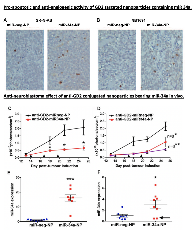
Here’s what makes this study so exciting: the team developed a nanoparticle system coated with anti-GD2, a molecule that recognizes and binds to GD2, a marker commonly found on neuroblastoma cells. Think of these GD2-coated nanoparticles as specialized delivery trucks with a precise address—they’re designed to deliver miR-34a.
Now, let’s dive into the details of miR-34a’s role. MiR-34a isn’t just any therapeutic agent—it’s a master regulator capable of influencing multiple genes involved in cell growth, survival, and blood vessel formation. By releasing miR-34a into tumour cells, this study activated pathways that induced cell death and suppressed angiogenesis, preventing the tumour from forming new blood vessels. It’s almost as if miR-34a is a conductor orchestrating a complex, multi-step attack on cancer, using the tumour’s own cellular mechanisms against it.
The Results? A Direct and Multi-Layered Attack on Tumor’s
In their mouse model, the GD2-targeted nanoparticles packed with miR-34a significantly reduced tumour growth. These “smart” nanoparticles didn’t just shrink tumors by inducing apoptosis (cell death); they also cut off the tumor’s blood supply by promoting the expression of TIMP2, an anti-angiogenic protein. Essentially, the tumor cells were directly targeted and deprived of the resources they needed to survive—a powerful one-two punch.
Where Do We Go From Here?
This study is an excellent example of how targeted therapies could evolve to tackle other types of cancer. Traditional therapies, like chemotherapy, often affect both healthy and cancerous cells, leading to significant side effects. In contrast, this targeted approach delivers miR-34a specifically to neuroblastoma cells, which could be especially beneficial for pediatric patients who need treatments that minimize harm to developing bodies. Imagine pairing nanoparticles like these with different therapeutic targets, such as GPC2, ALK, or PDL1, or even combining them with existing treatments to boost effectiveness while minimizing side effects. For those in the field, the potential here feels like a breakthrough waiting to happen.
Neuroblastoma is particularly challenging to treat, especially when tumours become resistant to chemotherapy. This resistance is compounded by tumour heterogeneity—these cancers comprise different cell types, specifically adrenergic and mesenchymal cells. This variability affects treatment responses and plays a role in metastasis and how aggressively the cancer can spread.
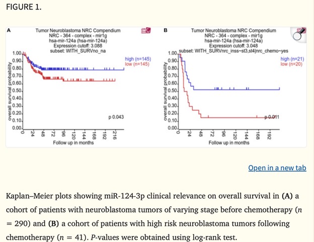
MicroRNAs (miRNAs) are small RNA molecules that regulate gene expression, and miR-124-3p has emerged as a promising player in cancer research. A Kaplan–Meier plot in the study (Figure 1) shows a strong association between low miR-124-3p levels and poorer survival rates in neuroblastoma patients, underscoring its potential impact on patient outcomes.
Our group’s study specifically examined how miR-124-3p might help reverse chemotherapy resistance and inhibit tumour cell growth in neuroblastoma. Excitingly, it has the potential to reduce cancer cell survival and increase their sensitivity to chemotherapy—an important breakthrough for treating resistant neuroblastomas.
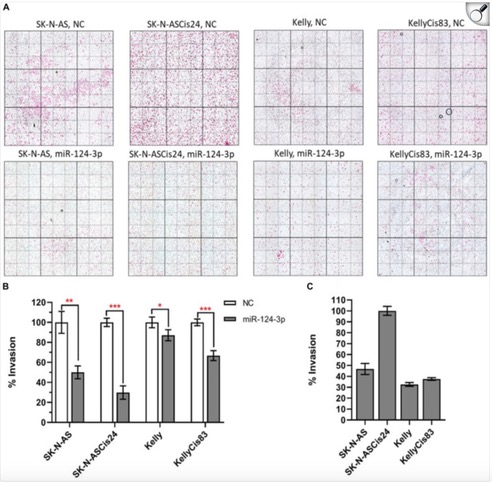
The study found that miR-124-3p directly targets genes involved in the epithelial-to-mesenchymal transition (EMT), a process that makes cancer cells more invasive and treatment-resistant. By suppressing these genes, miR-124-3p can reverse EMT, shifting cells to a less aggressive, more treatment-sensitive state. Our group observed that increased miR-124-3p significantly reduced neuroblastoma cell invasion (Figure 2). In SK-N-AS cells and their drug-resistant form, invasion dropped by 50% and 70%. In Kelly cells and their resistant form, invasion decreased by 10% and 30%. The most invasive of all, the drug-resistant SK-N-ASCis24 cells, showed the most substantial decrease in invasion after miR-124-3p treatment. This suggests that miR-124-3p could help limit neuroblastoma spread, highlighting its therapeutic potential.
While miR-124-3p isn’t part of my project, seeing how different molecular mechanisms can be harnessed to develop cancer therapies is always inspiring. Using miRNAs to sensitize resistant cancer cells to treatment could complement approaches like immunotherapies or vaccines, like the one I’m working on. Understanding these molecular pathways brings fresh perspectives on weakening cancer cells and making treatments more effective.
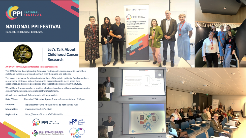
The RCSI Cancer Bioengineering Group hosted an in-person event during the National PPI Festival 2024 to share their childhood cancer research and connect with the public and patients.
We welcomed members of the public, family members of children with cancer, researchers, clinicians, and patient/community organisations on October 17th. Our past lab members and students paid a visit, too! Our group shared ongoing research on neuroblastoma biology and finding new treatments. Prof Cormac Owens from CHI brought us through the journey of clinical trials in neuroblastoma patients. We heard the heartbreaking story of the brave young man who lost his life to neuroblastoma and his parents who never gave up. This truly inspirational family founded a charity – the Conor Foley Neuroblastoma Cancer Research Foundation, to support curiosity-driven and translationally-focused research. The Foleys know very well how important it is to return happy days to kids and their families.
Cancer is the 2nd most common cause of death among children after accidents.
Childhood cancer is an umbrella term for many other types of this disease. Every September, many charities, researchers and parents of children with cancer work hard to raise awareness of this cancer. You may learn more about kids with cancer, their loving families, the doctors and caregivers who look after them and treat them, the young survivors of cancer and those kids and teens who lost their battle, and the scientists who work hard to find a way to stop childhood cancer.
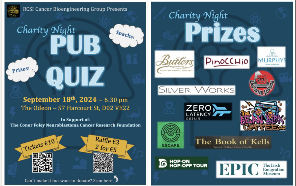
This year, our research team will run the Pub Quiz on September 18th, 2024, in honour of Childhood Cancer Awareness Month. All donations will go to the Conor Foley Neuroblastoma Research Foundation (CFNRF).
If you would like to get involved in this amazing challenge and help us raise vital funds for childhood cancers, you can contribute to our fundraising page:
HDAC inhibitors are drugs that target specific enzymes involved in gene regulation. This study tested broad-spectrum HDAC inhibitors as standalone treatments and combined them with doxorubicin, a well-known chemotherapy drug.
But why on Zebrafish? The zebrafish model provides a rapid and efficient means of testing these treatments, offering valuable insights into their potential use in combating neuroblastoma. This model allows for assessing drug efficacy and helps understand the associated toxicities quickly, making it a powerful tool for developing new anti-cancer therapies.
In the study, fish larvae were implanted with fluorescently labelled, well-established neuroblastoma cell line (SK-N-BE(2)-C) and patient samples (HD-N33, NB-S-124) to grow tumours. Non-cancerous cells (VH7 fibroblasts) were utilized to verify that tumour progression in zebrafish was specific to tumour cells. The engraftment of human cells into fish larvae was confirmed by immunohistochemistry (IHC) staining on zebrafish sections injected with neuroblastoma cells (SK-N-BE). This was achieved using a STEM121 antibody that reacts specifically with a human cytoplasmic protein. The findings showed that pediatric tumour cells survive and grow in the zebrafish model at rates like those observed in human tumours.
Before testing drug efficacy in zebrafish xenografts, optimal drug concentrations and maximal tolerated doses (MTD) were determined. Toxicity tests were conducted by treating fish larvae cells for three days without tumour cell injection to identify the maximum tolerated dose that did not cause observable morbidity, changes in morphology, or severe aberrations in larval behaviour. and lethal dose (LD) for each compound. To find optimal drug concentrations, larvae with xenografted tumour cells were incubated with increasing drug doses 24 hours post-implantation to the maximally tolerated dose (MTD). The relative IC50 values were then determined based on changes in tumour mass volume.
To evaluate the treatment, SK-N-BE(2)- cells were used to test the broad-spectrum HDAC inhibitors, including panobinostat, vorinostat, and tubastatin A, both alone and combined with doxorubicin. The partial response rate (PR) was measured to see how well different drug combinations work to shrink tumours in the zebrafish model. Here’s what they found: Doxorubicin combined with panobinostat resulted in a 23% PR, Doxorubicin combined with tubastatin A showed a 31% PR, and Doxorubicin combined with vorinostat achieved the best result with a 36% PR.
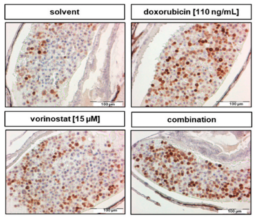
To test the effectiveness of the HDAC inhibitor treatment, they monitored the tumour growth using a confocal microscope before and 48 hours after giving the drug. The test revealed that a 48-hour treatment of SK-N-BE (2)-C zebrafish xenografts with vorinostat and doxorubicin alone, `and in combination, increased cell death. The combination of these two drugs was the most effective, causing a significant increase in cancer cell death (apoptosis) by decreasing cell proliferation, as indicated by reduced PPH3 marker and activating the number of Cleaved caspase-3 (Figure 1).
Figure 1: Treatment for 48 h with Vorinostat, doxorubicin, or a combination of both increased the amount of cleaved caspase-3 and reduced mitotic tumour cells. Adapted from Pharmaceuticals2020, 13(11), 345
In essence, this study validates the use of HDAC inhibitors in treating neuroblastoma and paves the way for broader applications of zebrafish models in cancer research. As we look to the future, these innovative models could significantly enhance our ability to develop effective cancer therapies, making strides towards better treatments and, ultimately, more effective cures.
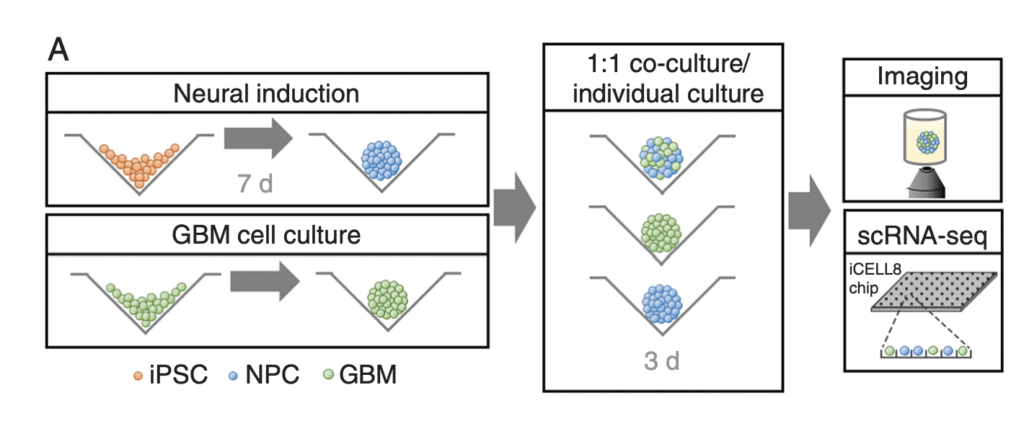
This article by Krieger et al. discusses the most common form of brain cancer called glioblastoma. Due to its highly aggressive nature, research must be conducted consistently and rapidly to develop new treatments. This has proven challenging due to primary tumours being resected before further research can be done, as well as the lack of current technologies to fully explore relationships between GBM and surrounding brain tissues. This study aimed to study the aforementioned interactions in under 4 weeks, accounting for the rapid progression of the disease in real life.
GBM cells were first derived from four patients and treated with glutamine, heparin, epidermal and fibroblast growth factors, then underwent a sequence of manipulations, such as second-generation replication lentivirus infection of GBM cells, iPSC line 409b2 inoculation in Aggrewell plates and later manipulation with invasion assays, and scRNA sequencing, which, along with the Aggrewell cells, produced neural progenitor cell spheroids for analysis. Confocal microscopy and the developed image processing algorithm allowed for visualization of these cells following fluoroscopy and depicted consistent growth of tumour cells. There was also the growth of microtubules. Any dissociated organoids were then co-cultured with GBM cells again, promoting interaction between the two. Further analysis revealed the upregulation of 45 genes, including PAX6, GJA1, GPC3, and others involved in cell regulation.
In conclusion, this novel mechanism of analysis of GBM cells using Aggrewell plates provided fruitful results, indicating intricate relationships between GBM cells and organoids, providing crucial insight for treatments by elucidating specific gene expression, heterogeneity of cells, and offering new targets based on ligand-receptor interactions. The particular relevance of this study to my work is regarding the usage of Aggrewell plates, which I am currently studying to determine how best to keep cells growing successfully within the wells. This article proves the usability and efficiency of Aggrewell and establishes its crucial role in the realm of brain cancer treatment research.
Hi there, Federica here! In the fast-paced world of scientific research, staying informed about the latest studies and breakthroughs is crucial. It enables researchers to build upon existing knowledge, avoid redundant efforts, and discover new directions for their work. That’s why we’ve started a new series of blog posts highlighting recent papers and explaining their significance for our research.
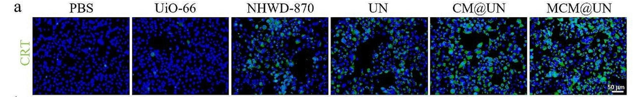
The researchers investigated a combination of immune checkpoint blockade (ICB) and other drugs to turn “immune-cold” tumours (which evade the immune system) into “immune-hot” tumours (which the immune system can attack). They developed a special delivery system using nanoparticles called metal-organic frameworks (MOFs). These nanoparticles were loaded with two types of drugs—a TLR7/8 agonist and an epigenetic inhibitor (BRD4 inhibitor). To make the nanoparticles even more effective, they were coated with vesicles from the cancer cells themselves. This coating helps the nanoparticles specifically target cancer cells.
But how does it work?
The nanoparticles are designed to find and enter triple-negative breast cancer (TNBC) cells. Once inside, the drugs prompt the cancer cells to break apart and release signals that alert the immune system. These signals attract dendritic cells, which then activate CD8+ T cells—the body’s natural cancer fighters. The TLR7/8 agonist further enhances this immune response, making the treatment more powerful.
In both laboratory tests and animal models, this method showed significant promise. It not only slowed down tumour growth but also improved the body’s immune response to cancer. Importantly, the study found that this approach could remodel the tumour environment, making it more hostile to cancer cells. For example, they wanted to verify that their combined delivery system could really boost the body’s ability to fight tumours. They focused on a protein called calreticulin (CRT) that, when it shows up on the surface of tumour cells, helps the immune system spot and remove them. They found that when they used their special delivery system (CM@UN and MCM@UN), the levels of CRT on the surface of tumour cells went way up. This was especially true for the MCM@UN group, showing just how powerful their method was in getting the immune system to attack the tumours.
The original image was published in J Nanobiotechnology. 2024; 22: 296.
So, why is this study important for my work?
The principles of enhancing the immune system’s ability to fight cancer are central to both the research in the study and in my project. Like the nanoparticles in the study, mRNA vaccines can be designed to specifically target cancer cells, ensuring that the treatment reaches its intended destination. Another similarity is how the drugs activate the immune system, which parallels how mRNA vaccines work—by training the immune system to recognise and attack cancer cells.
I find this study really interesting as it sheds light on innovative strategies for cancer treatment and provides valuable insights that can inform and inspire our research on developing mRNA vaccines for childhood neuroblastoma!
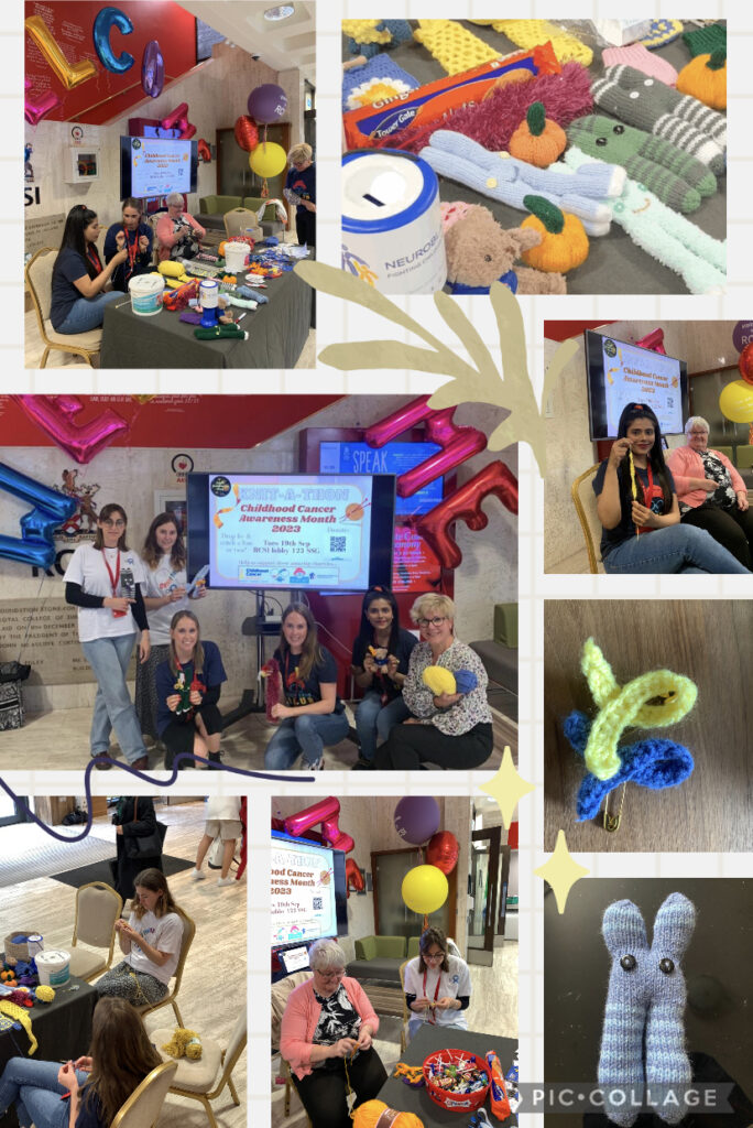
A wonderful day of knitting – Knit-A-Thon-2023 raised 913 euros. A massive thank you to everyone who stopped by and donated on the day and beyond. Every cent counts! The money was split evenly between our four chosen charities: The Conor Foley Neuroblastoma Research Foundation (CFNRF), Neuroblastoma UK (NBUK), Oscars Kids and Childhood Cancer Ireland (CCI). These charities were established and are run by parents, some of whom lost their children to cancer. They continue their children’s legacy, doing an amazing job of advocating for children with cancer and better funding for research and aftercare.
Knit-A-Thon 2023
And a special thank you to Ciara’s mam Aggie for the amazing handmade raffle prizes (chromosomes, antibodies, cup holders and many more) and a Master class on the day! We thank Jenny Duffy (RCSI Events and Communications Coordinator) for her time crocheting with us and for us! Thanks to Anggie’s and Jenny’s skills, there were lots of mascots to win – and many of them collected already. We much appreciate the support from the RCSI Estates and Porters who looked after us on the day.