#JournalClubwithFederica:How small RNAs contribute to neuroblastoma biology

We’ve recently started a new journal club series focusing on papers published by our research group over the past few years. The paper I chose is titled “A Context-Dependent Role for MiR-124-3p on Cell Phenotype, Viability and Chemosensitivity in Neuroblastoma in vitro“. It explores the anti-cancer potential of miR-124-3p in neuroblastoma.

Neuroblastoma is particularly challenging to treat, especially when tumours become resistant to chemotherapy. This resistance is compounded by tumour heterogeneity—these cancers comprise different cell types, specifically adrenergic and mesenchymal cells. This variability affects treatment responses and plays a role in metastasis and how aggressively the cancer can spread.

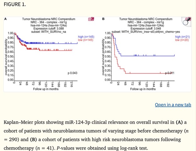
MicroRNAs (miRNAs) are small RNA molecules that regulate gene expression, and miR-124-3p has emerged as a promising player in cancer research. A Kaplan–Meier plot in the study (Figure 1) shows a strong association between low miR-124-3p levels and poorer survival rates in neuroblastoma patients, underscoring its potential impact on patient outcomes.

Our group’s study specifically examined how miR-124-3p might help reverse chemotherapy resistance and inhibit tumour cell growth in neuroblastoma. Excitingly, it has the potential to reduce cancer cell survival and increase their sensitivity to chemotherapy—an important breakthrough for treating resistant neuroblastomas.

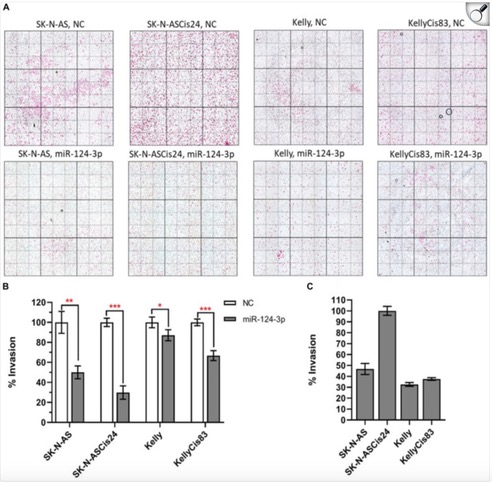
The study found that miR-124-3p directly targets genes involved in the epithelial-to-mesenchymal transition (EMT), a process that makes cancer cells more invasive and treatment-resistant. By suppressing these genes, miR-124-3p can reverse EMT, shifting cells to a less aggressive, more treatment-sensitive state. Our group observed that increased miR-124-3p significantly reduced neuroblastoma cell invasion (Figure 2). In SK-N-AS cells and their drug-resistant form, invasion dropped by 50% and 70%. In Kelly cells and their resistant form, invasion decreased by 10% and 30%. The most invasive of all, the drug-resistant SK-N-ASCis24 cells, showed the most substantial decrease in invasion after miR-124-3p treatment. This suggests that miR-124-3p could help limit neuroblastoma spread, highlighting its therapeutic potential.

While miR-124-3p isn’t part of my project, seeing how different molecular mechanisms can be harnessed to develop cancer therapies is always inspiring. Using miRNAs to sensitize resistant cancer cells to treatment could complement approaches like immunotherapies or vaccines, like the one I’m working on. Understanding these molecular pathways brings fresh perspectives on weakening cancer cells and making treatments more effective.

Written by Federica Cottone

#JournalClubwithEve: Unraveling Neuroblastoma Metastasis – My Exciting PhD Journey into 3D Models

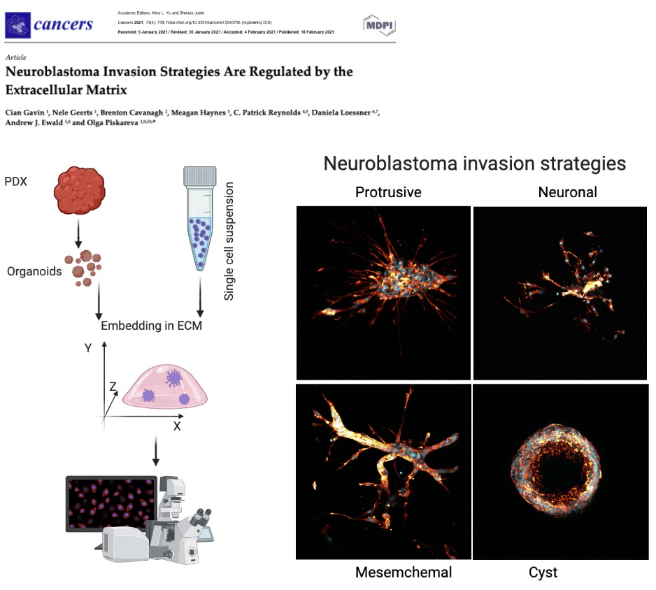
As a new PhD student, I’m incredibly excited to dive into cancer research, and what better way to kick off this journey than by exploring 3D models to study neuroblastoma metastasis? Neuroblastoma is one of the most common childhood cancers, and about 50% of patients have metastatic disease at diagnosis. Understanding how these cells spread is key to developing better therapies, which is why this recent study by Gavin et al. (2021) caught my eye.

So, what did the researchers do? They used something called patient-derived xenografts (PDX) and cell lines to grow organoids (tiny mini-tumors) in a 3D extracellular matrix (ECM). This ECM mimics the environment these cells would encounter in the body, which is super important because cells behave very differently in 3D than in the typical 2D Petri dishes. It’s like giving the cells an entire landscape to explore rather than just a flat road—suddenly, they have mountains to climb and valleys to cross, allowing them to behave much more like they would inside the body!

One of the coolest things about this study is how the neuroblastoma cells developed various invasion strategies based on their environment. Some stayed in tightly knit groups, while others decided to go full-on lone wolf, sending out long, thin projections to explore the surrounding matrix. These cells are smart-adapting to different ECM compositions like Matrigel (which is rich in laminin and collagen), made them change their behaviour entirely. It’s like they’re navigating an obstacle course, with each new challenge requiring a different tactic!

Let’s Talk Actin Filaments!

Now, this is where it gets super cool (and nerdy in the best way!). The images captured by confocal microscopy are stunning. They show actin filaments—the internal skeleton of the cells—as they help the cancer cells move and invade new areas. The actin filaments form these amazing, intricate networks that shape the cells and allow them to stretch and invade. It’s almost like watching tiny construction workers build bridges and tunnels as they move forward. Check out this confocal image showing the red filaments—how awesome is that?!

Written by Eve O’Donoghue

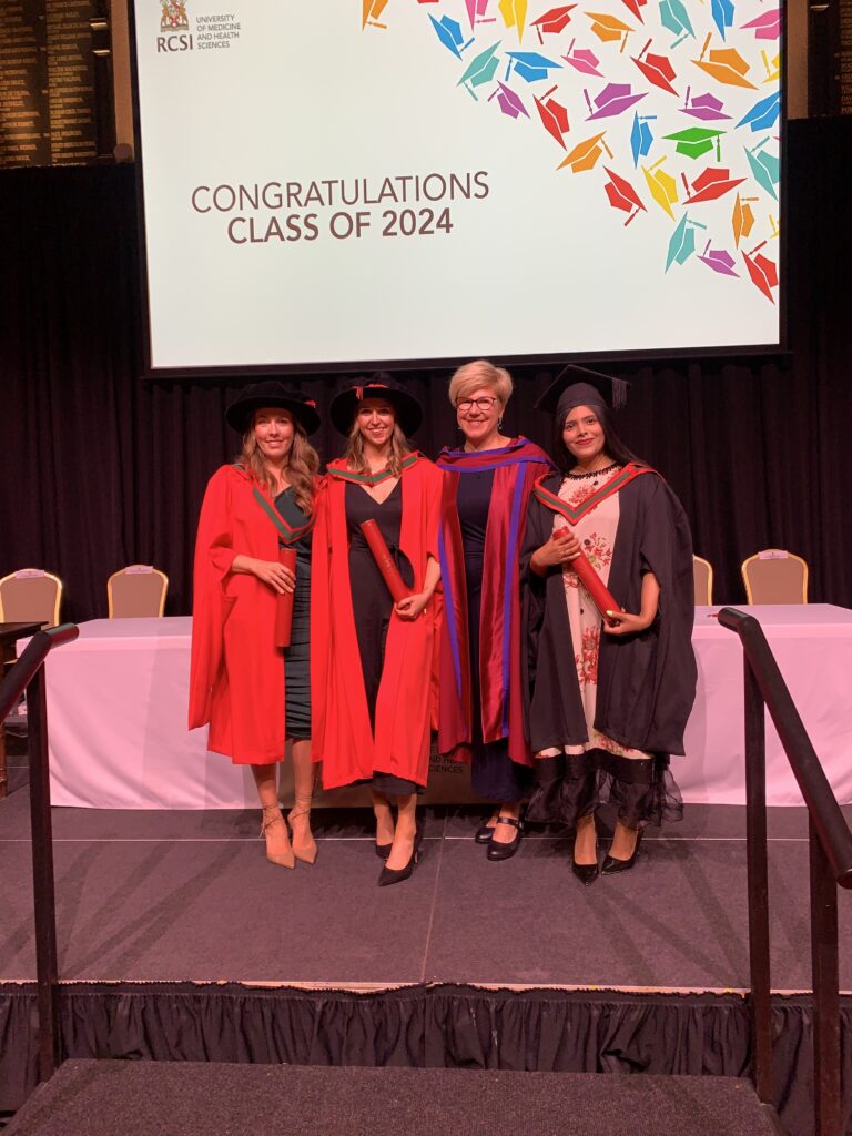
Class 2024: Congratulations to Ciara, Ellen and Rabia!

Massive congratulations on the official moulding of PhD and MSc by Research to our promising young scientists: Rabia Saleem, Dr Ciara Gallagher and Dr Ellen King! Great accomplishments!

Three different journeys, with two through the COVID-19 pandemic. The full range of ups and downs. Who said that the PhD is a straight line? It has never been. It is more like the Irish weather: some days are sunny and bright, and some have scattered showers, gale winds and stormy snow, with sunshine developing elsewhere. The journey was spiced up with publications, conferences, travels, days out and fundraising events with the team.

It is a proud moment for me as well. 🙂 Three PhD and one MSc by Research students graduated within the last 12 months.

Of note, Ellen was behind our Twitter activities in the past, making our team visible!

Wish you all the best of luck on your new adventure!

Olga Piskareva

Congratulations to a new Dr in the house: Dr Ellen King

Huge congrats to a newly minted Dr Ellen King!  She passed her PhD viva on April 9. This is a testimony to your dedication, strong will and hard work. May this PhD be the beginning of many more successful endeavours, Ellen!

We thank examiners Prof Sally-Ann Cryan (RCSI) and Prof Joanne Lysaght (TCD) for the time and expertise they provided.

We also thank the RCSI PhD Programme for their generous support!

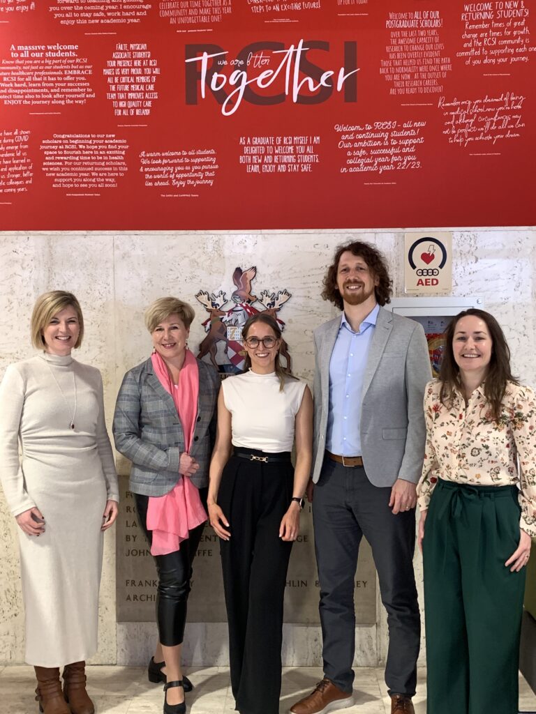
From left to right: Prof Joanne Lysaght, Dr Ellen King, Dr Olga Piskareva & Prof Sally-Ann Cryan

How things work in science: Scaffolding

At the Cancer Bioengineering Group, we use different types of scaffolds to mimic the 3D structure of tumours outside the body. We use these scaffolds to test new therapeutics and understand the tumour microenvironment. But I bet you didn’t think we had this in common with spiders?

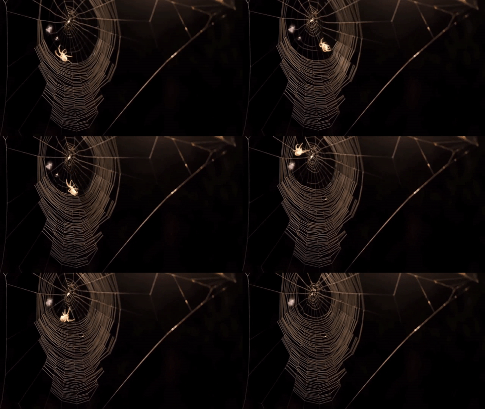
Spiders make their webs by producing silk from specialized glands in their abdomen. They release the silk through spinnerets located at the back of their abdomen, then use their legs to manipulate the silk strands into intricate patterns, depending on the species and purpose of the web.

The process of web building begins with a scaffold. The specialized glands that spiders use are called spinnerets, and they produce liquid silk proteins that solidify into a thread when they come into contact with air. Using their many legs, spiders can manipulate the threads by changing the speed and tension they enforce on the silk, thus controlling thickness, stickiness and strength. They first lay a framework of non-sticky threads, known as scaffolding. And layer by layer, different species of spiders will add their own artistic sticky silk design to the scaffold depending on their aim. Take the deadly redback spider for example, these guys have a utilitarian approach to web building relying on their webs mainly for shelter and capturing prey. As such, they don’t put much effort into producing irregular and messy homes. In comparison, the orb-weaving spider produces “Mona Lisa”-like designs, with complex geometric patterns and intricate designs. The differences in effort seem to come from the environments in which the webs are located, with the redbacks choosing more sheltered environments and thus not needing much strength to their webs. Whereas orb-weaving spiders are more adapted to a range of environments, from forests to grasslands to urban gardens. So, while the redback gets a lot of attention for their neurotoxic venom, they need to step up their artistic skills to match that of their orb-weaving colleagues.

The redback spider and its webs are reminiscent of an aggressive tumour, which is erratic, dangerous, and unpredictable. We want to find the “anti-venom” for such tumours so we can wipe them out for good.

Watch this amazing web-building timelapse by BBC Earth.

Written by Ellen King

Congratulations to Dr Ciara Gallagher!


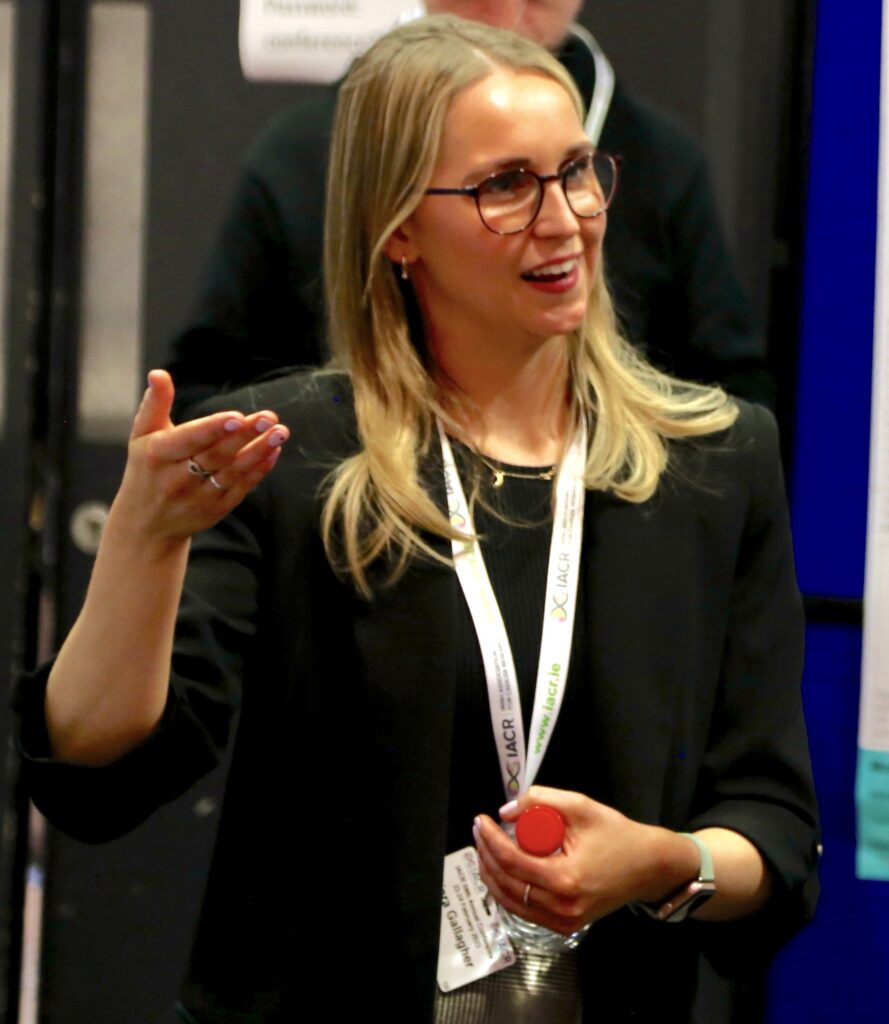
Huge congrats to a newly minted Dr Ciara Gallagher!  She defended her PhD on March 8 – International Women’s Day. Your enthusiasm and perseverance are truly fascinating! May this be the stepping stone towards a brighter future, Ciara!

We thank examiners Dr Marie McIlroy (RCSI) and Prof Jan Škoda (Masaryk Uni) for the time and expertise they provided.

We also thank the Irish Research Council for their generous support!

Dr Ciara Murphy (Chair), Dr Olga Piskareva (Supervisor), Dr Ciara Gallagher, Prof Jan Skoda (examiner), Dr Marie McIlroy (Examiner)

Growing cancer cells in 3D

Hi there, Ciara here again, a final-year PhD student in our research group. I can’t believe September has rolled around again, meaning one thing: it’s Childhood Cancer Awareness Month (CCAM). In honour of this month, I would like to tell you a little bit about the childhood cancer we study in our lab and the research that I do to one day help save children from this disease. 

Neuroblastoma is an aggressive childhood cancer, with sadly only 20% of late-stage patients surviving after 5 years. Progressive disease and cancer relapse are common in neuroblastoma. This is due to standard treatment regimens not being adequate for treating high-risk patients. Current treatment also may cause a series of adverse reactions in patients. Therefore, my research focuses on developing a 3D model of high-risk neuroblastoma that models the cancer more accurately in a laboratory setting. This will act as a beneficial platform to test whether new therapies effectively fight the patients’ cancer cells, leading to better treatment options for children with neuroblastoma.  

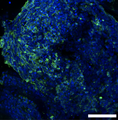
Below is a picture of how we grow these cancerous cells on our 3D model and visualise them with fluorescent stains. When we can see them like this under a microscope, we can study how they move and grow to help us understand how to treat them. 

Here, we can see the cells growing on our 3D cancer model. This image is magnified by 200 times to be able to see the individual cancer cells. The green stain is the outside of our cancer cells, or we use the term, the cell membrane. The blue is the inside, or as some of you may know the term, the nucleus of the cell.   (It is amazing what we can see with the power of microscopes, right?) 

As you may know, every year, we support amazing charities by raising vital funds to keep the fight against childhood cancer going. Keep your eyes peeled on our Twitter for updates on what crazy activity we have committed to this year!!  

Written by Ciara Gallagher

Childhood Cancer Awareness Month 2023

Every September, we celebrate Childhood Cancer Awareness Month. This is a great opportunity to raise awareness about childhood cancer. Unfortunately, kids get cancer, too. While much research has been done to understand how cancer develops in adults, we still know very little about what exactly leads to cancer in children.

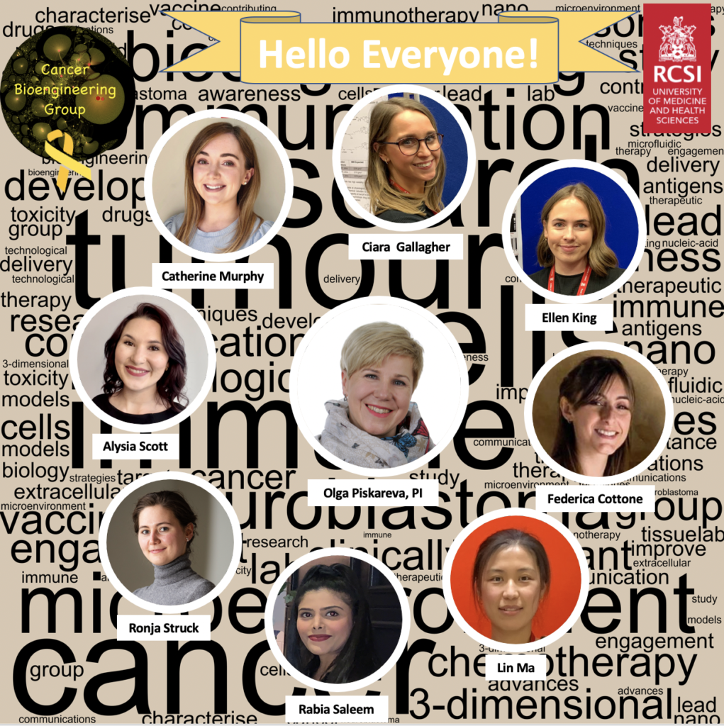
We are the Cancer BioEngineering Group led by Dr Olga Piskareva at the RCSI University of Medicine and Health Sciences. Our research focuses on neuroblastoma, an aggressive childhood cancer of immature nerves. The group has 7 PhD students developing research projects around neuroblastoma biology. One postgraduate student successfully defended her work and was awarded a PhD last month.

We are a dynamic group proud to be engaged in research, science communication and patient involvement. We do that through different initiatives. Throughout September, we will share many of them and invite you to keep following us on social media. 

Team 2023

Our projects address topics related to neuroblastoma microenvironment, cell interactions, tumour resistance and the development of new therapies. To do that, we use 3D in vitro models, identify immunotherapeutic targets and evaluate extracellular vesicles.  

We are always happy to answer questions and interact with the public. Follow us on our social media channels and read our blog to learn more about us and our research.  

We are running a fundraising event, “A knit-a-thon,” on the 19th of September. Stay tuned!

Thanks for reading, and we go ahead with neuroblastoma research! 

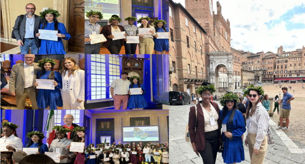
Experiencing Elegance: A Graduation Ceremony at the University of Siena, Italy

This blog takes you to the exciting scene of my MSc graduation ceremony at the University of Siena, Italy, completed with the prestigious laurel wreath.

I graduated during COVID-19, but there was no graduation ceremony at that time. Years later, I was invited to attend an “Alumni conference” by the University of Siena, but the plan was still unclear. When we arrived in Siena, we came across that there was a convocation ceremony tomorrow. Hold on! What? Yes, after years of waiting, it was finally taking place on June 7, 2023.

The next morning, all the former students from 48 countries came together in the University’s Grand Piazza del Duomo, where all of the Professors and sponsors, robed in their academic attire, delivered speeches that inspired and reminded us of the responsibility that comes with education, which ended in the most captivating moment of adorning us with laurel wreaths stating that “Rating your thesis attributes by authority granted to me by director I confer you the Masters Diploma in Vaccinology and Drug Development, Congratulations!”. The weight of this academic success was alleviated by our family members’ joyful yells and applause.

Walking out of the ceremony, wreathed in laurel, walking through Siena’s streets with classmates I’ve never met in person, hearing these words “Complimenti! Felicitazioni!” from commoners, I came back to Dublin with an ethereal sensation of pride and belonging that will remain with me for life. Altogether, It was a once-in-a-lifetime experience for me.

Here are some glimpses of the ceremony:

P.S.: But this was not the end. I embarked on a wet-lab MSc in RCSI Dublin. As I am typing these lines, my MSc by Research work has just been submitted for examination, marking another hallmark and opening a new chapter in my life, “the PhD journey”. The new chapter – the new challenges and opportunities!

Written by Rabia Saleem

A newly minted Dr. Catherine Murphy

Huge congrats to a newly minted Dr Catherine Murphy! ✨ She successfully defended her PhD work yesterday. Hard work and dedication paid off. Well done, Catherine!

We thank examiners Drs Oran Kennedy and Niamh Buckley for their time and expertise provided.

We also thank Neuroblastoma UK for their generous support!