How things work in science: Gene editing technology

Few advancements in biomedical sciences hold as much promise for revolutionising cancer research as CRISPR-Cas9. This ground-breaking gene-editing tool has sparked a wave of innovation, offering precision and efficiency in manipulating the human genome in the fight against cancer.

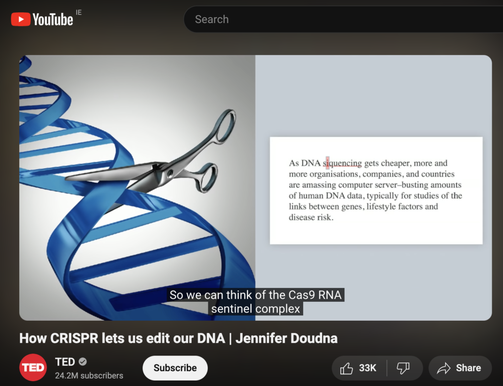
Now, what is it? CRISPR is basically an acronym for a very long name Clustered Regularly Interspaced Short Palindromic Repeats Associated Protein 9 or CRISPR-Cas9 for short. It was found in simple organisms such as archaea and bacteria. Interestingly, this is a component of bacterial immune systems that can cut DNA. So, this feature was proposed for use as a gene editing tool, a kind of precise pair of molecular scissors that can cut a target DNA sequence. So, the CRISPR-Cas9 scissors allow us to precisely edit the DNA sequence of living organisms by adding in (knock-in) or removing (knockout) a gene of interest.

For cancer research, for example, the CRISPR-Cas9 scissors can be used to introduce therapeutic genes or correct mutations associated with cancer predisposition syndromes. Meanwhile, those scissors can also disrupt genes involved in treatment resistance, sensitising cancer cells to existing therapies.

Jennifer Doudna and Emmanuelle Charpentier have won the 2020 Nobel Prize in Chemistry “for the development of a method for genome editing.”. A nice accompanying piece was published in The Conversation, highlighting the history of these scissors and the politics behind it.

Jennifer Doudna explains this revolutionary genetic engineering tool in a TED lecture. However, she warns:

“All of us have a huge responsibility to consider carefully both the unintended consequences as well as the intended impacts of a scientific breakthrough.”

I hope you enjoyed it!

Witten by Rabia Saleem
Congratulations to a new Dr in the house: Dr Ellen King

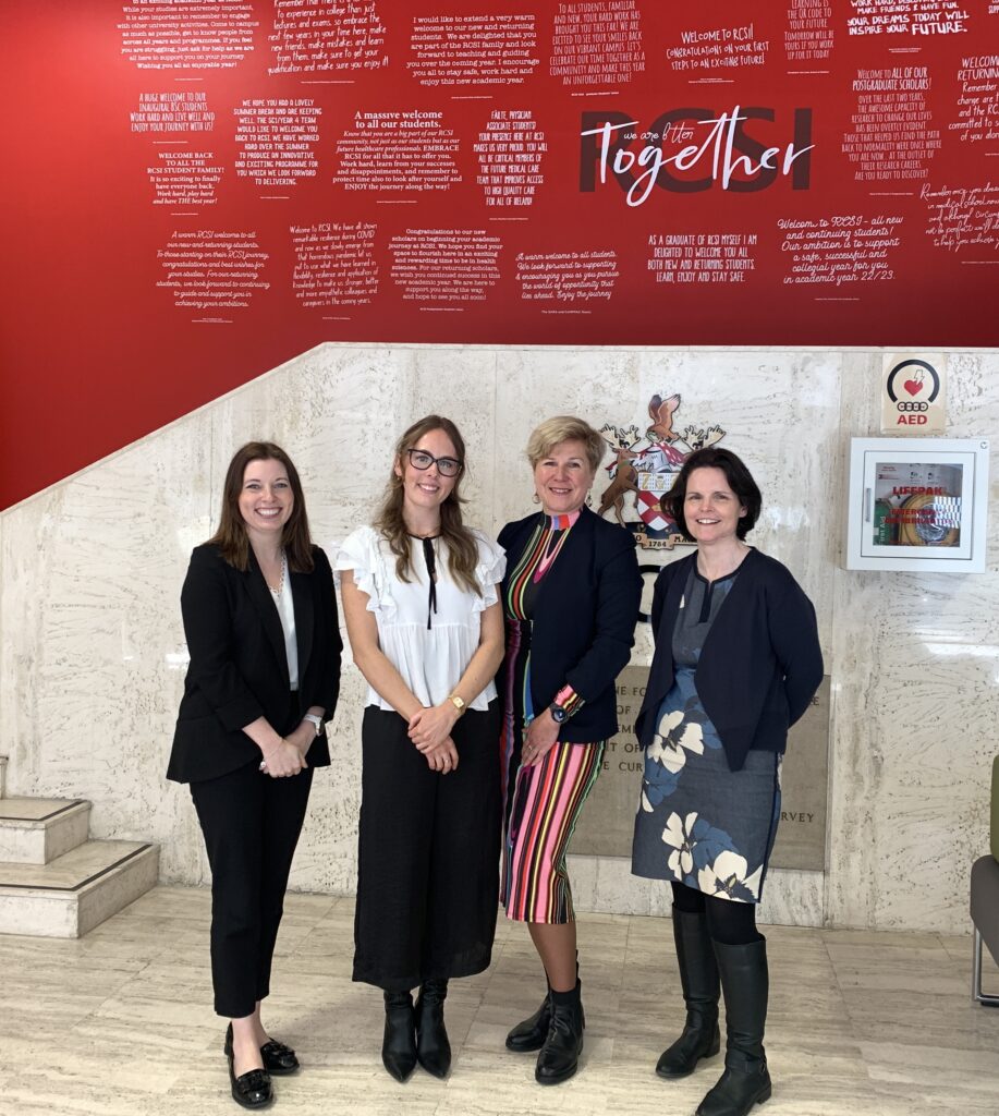
Huge congrats to a newly minted Dr Ellen King!  She passed her PhD viva on April 9. This is a testimony to your dedication, strong will and hard work. May this PhD be the beginning of many more successful endeavours, Ellen!

We thank examiners Prof Sally-Ann Cryan (RCSI) and Prof Joanne Lysaght (TCD) for the time and expertise they provided.

We also thank the RCSI PhD Programme for their generous support!

From left to right: Prof Joanne Lysaght, Dr Ellen King, Dr Olga Piskareva & Prof Sally-Ann Cryan

How things work in science: targeting cell components.

How do researchers study cells? How do we get the nitty gritty?

We use many methods to tag and chase various cell components. One of my favourites is fluorescent microscopy. It allows the use of nearly all spectrum of colours from blue to purple in one go. However, we prefer to narrow it down to 2-3 colours and avoid their overlap.

How does it work? First, we use DAPI or Hoescht, which are blue fluorescent dyes used to stain DNA. This way, we tag the nucleus of the cell. Then, we tag a protein of interest. In our case, it was MYCN, a protein that acts as a transcription factor. MYCN amplification is associated with poor prognosis in neuroblastoma. As a transcription factor, it binds to genomic DNA and is located in the nucleus. We used a specific antibody that was labelled with a green fluorescent dye. Look at the image below. The green colour pattern overlaps with the blue colour. Then, we tagged the cytoskeleton, a complex of various proteins that hold the cell architecture and dynamics. We used phalloidin with red fluorescence. It is a highly selective bicyclic peptide and a popular choice for staining actin filaments.

Neuroblastoma organoids stained with DAPI, Phalloidin and anti-MYCN antibody. This work was done during the Fulbright journey to Ewald’s Lab at Johns Hopkins

Now, we can enjoy visualising cells and test different research questions. For example, how do cells respond to a drug? Or how do neuroblastoma cells spread?

Written by Olga Piskareva

How things work in science: Scaffolding

At the Cancer Bioengineering Group, we use different types of scaffolds to mimic the 3D structure of tumours outside the body. We use these scaffolds to test new therapeutics and understand the tumour microenvironment. But I bet you didn’t think we had this in common with spiders?

Spiders make their webs by producing silk from specialized glands in their abdomen. They release the silk through spinnerets located at the back of their abdomen, then use their legs to manipulate the silk strands into intricate patterns, depending on the species and purpose of the web.

The process of web building begins with a scaffold. The specialized glands that spiders use are called spinnerets, and they produce liquid silk proteins that solidify into a thread when they come into contact with air. Using their many legs, spiders can manipulate the threads by changing the speed and tension they enforce on the silk, thus controlling thickness, stickiness and strength. They first lay a framework of non-sticky threads, known as scaffolding. And layer by layer, different species of spiders will add their own artistic sticky silk design to the scaffold depending on their aim. Take the deadly redback spider for example, these guys have a utilitarian approach to web building relying on their webs mainly for shelter and capturing prey. As such, they don’t put much effort into producing irregular and messy homes. In comparison, the orb-weaving spider produces “Mona Lisa”-like designs, with complex geometric patterns and intricate designs. The differences in effort seem to come from the environments in which the webs are located, with the redbacks choosing more sheltered environments and thus not needing much strength to their webs. Whereas orb-weaving spiders are more adapted to a range of environments, from forests to grasslands to urban gardens. So, while the redback gets a lot of attention for their neurotoxic venom, they need to step up their artistic skills to match that of their orb-weaving colleagues.

The redback spider and its webs are reminiscent of an aggressive tumour, which is erratic, dangerous, and unpredictable. We want to find the “anti-venom” for such tumours so we can wipe them out for good.

Watch this amazing web-building timelapse by BBC Earth.

Written by Ellen King

Congratulations to Dr Ciara Gallagher!


Huge congrats to a newly minted Dr Ciara Gallagher!  She defended her PhD on March 8 – International Women’s Day. Your enthusiasm and perseverance are truly fascinating! May this be the stepping stone towards a brighter future, Ciara!

We thank examiners Dr Marie McIlroy (RCSI) and Prof Jan Škoda (Masaryk Uni) for the time and expertise they provided.

We also thank the Irish Research Council for their generous support!

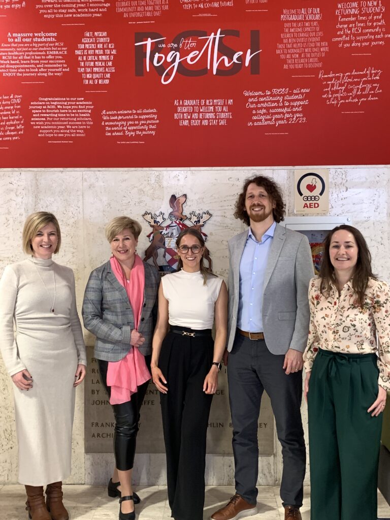
Dr Ciara Murphy (Chair), Dr Olga Piskareva (Supervisor), Dr Ciara Gallagher, Prof Jan Skoda (examiner), Dr Marie McIlroy (Examiner)

Ever wonder how scientists figure out a specific protein’s role in cancer?

Researchers use various methods, but I employ gene knockdown in my experiments. Basically, I use small RNA molecules that specifically target and degrade the mRNA of my gene of interest. This leads to a decrease in the corresponding protein levels, enabling me to observe the effects on neuroblastoma cell behaviour.

I feel a bit like Sherlock Holmes, you know? I’m selectively putting my suspect protein – the one I’m eyeing – under the spotlight to see how it’s pulling the strings on the cell’s behaviour. It’s like I’m in a cellular mystery, complete with a gene knockout magnifying glass 🔍🧬🕵

So, what I’ve been up to these past months is knocking down my protein and trying to find answers to the following questions:

Can neuroblastoma cells survive? And if not, how do they meet their demise? Do they go on a growth spree and start proliferating? Are they capable of migration? And here’s the twist – when my protein of interest takes a dip, do other proteins decide to change their expression levels?

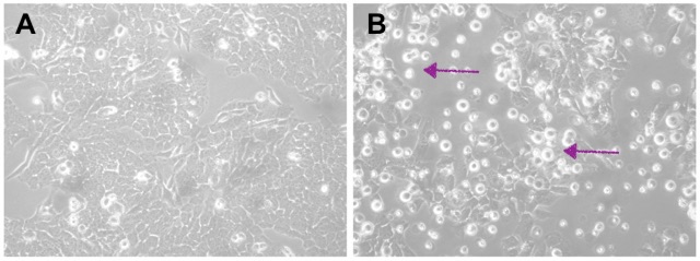
The picture below can probably help you get an idea of what I’ve done so far. Do you see those brighter spots in Pictures A and B? Those are dead cells. Their number indicates the proportion of dead cells after a treatment. Picture A has just a few; the majority are healthy and well-spread cells. This is our negative control, a condition when we show neuroblastoma cells that have been transfected, but no gene knockdown happened. Transfection is the term for introducing small RNA molecules. Now, in Picture B, when we knocked down the protein, it caused the death of the cells, and you can clearly see that from all those many little bright spots.

We have found answers to many of the previous questions, but new questions have arisen, and we can’t wait to answer them!

Written by Federica Cottone

International Childhood Cancer Day – 15 February 2024

We are celebrating #ICCD2024 with a Bake Sale and a Quiz. To earn a piece of cake, you have to answer a question correctly! Have a look at some:

  • Which civilisation first described cancer?
  • Where did the word cancer come from?
  • Do children get cancer?
  • What is the most common type of cancer in children?
  • Can the Human Papillomavirus (HPV) vaccine prevent cancer?
  • Can neuroblastoma begin to develop before birth?
  • What is the name of the nerve cell in which neuroblastoma begins to grow?
  • Can a child have a genetic predisposition to neuroblastoma?
  • What % stands for the incidence of neuroblastoma: 8 or 15?
  • What % stands for the neuroblastoma-related deaths: 8 or 15?
  • Does neuroblastoma first appear in the brain?
  • What does the letter N stand for in the gene MYCN?
  • How often does childhood cancer occur compared to adults?
  • How often does hereditary cancer happen in general?
  • Do you think that children are small adults when we talk about anticancer treatment?

How things work in Science: Classifiers

For our next little series introducing a different thing in science and how it works every week, I decided to focus on classifiers. With artificial intelligence becoming more and more prominent in our daily lives as of late, I thought this would be a good lead into the explicitly science-focused topics to come. So, what is a classifier? How does it work? And why does it matter?

At their core, classifiers are algorithms designed to categorize input data into predefined classes or categories. They learn patterns and relationships from labelled training data to make predictions on new, unseen data.

Once features are extracted, identified and quantified from labelled or annotated input data, mathematical models are employed for pattern recognition and predictions.

These models can range from simple decision trees to complex neural networks, each with its own strengths and weaknesses.

Training these models is an iterative process. That means to produce one good classifier, lots of classifiers were created in the process: Every time the pattern recognition is run, the annotated data is categorised by the classifier and compared to the annotation class. Prediction errors are corrected, and performance is optimised. This whole process is one iteration. How many iterations are required for a well-trained classifier varies widely and is largely dependent on the input data and application. For my tissue classifiers, it took up to 20,000 iterations.

Classifiers use these models to categorise unseen data into categories the user-defined at the start. In the figure, you can see my annotated histological slides from which the classifier extracted patterns to then classify the rest of the slide and entirely unseen slides into tumour (red), stroma (green) and background (blue) classes.

From identifying fraudulent transactions, filtering out junk mail, targeted advertising, and facial recognition to unlock your phone or diagnosing diseases, classifiers play a vital role in automating decision-making processes and driving advancements across a wide range of industries. Keep your eyes peeled, and you can find more classifiers in action all around you.

Written by Ronja Struck

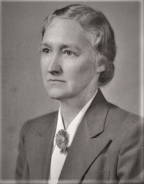
Women in Science: Dr. Margaret Jane Pittman

In the world of scientific research, Dr. Pittman’s name stands out. She was a bacteriologist and one of the founders of vaccines against infectious diseases such as typhoid, cholera, whooping cough and meningitis. Dr. Pittman achieved another incredible feat as the first woman to give rise to a National Institutes of Health Laboratory in the US. This accomplishment dispelled the persistent gender stereotypes that had long prevented women from pursuing careers in STEM and showcased her extraordinary credentials and experience.

Dr. Margaret Jane Pittman was born on January 20, 1901, and grew up in Prairie Grove, Arkansas. Dr Margaret Pittman graduated magna cum laude from Hendrix in 1923. In 1925, she took her first course in bacteriology. At the time, it was propitious. Later in the nineteenth century, bacteriology became a distinct biological science. She got her Ph.D. from the University of Chicago in 1929 and started working for the National Institutes of Health in 1936. She developed, assessed, and standardised immunisation programmes against cholera, whooping cough, typhoid, and other diseases during most of her career. She continued to work at NIH as an unpaid guest. She occasionally served as a consultant for the World Health Organisation and as a guest scientist in several countries, including Spain, Scotland, Egypt, and Iran.

Her work and relentless efforts have revolutionised the world of vaccines and immunisation. Dr Pittman’s Haemophilus influenzae study has significantly influenced the medical field. Based on her research, numerous lives have been saved due to the creation of potent vaccinations, which has led to a notable drop in diseases linked to Haemophilus influenza. Pittman’s collaborations with esteemed scientists like Drs. Jonas Salk and Albert Sabin were essential in creating vaccinations against polio. They collaborated to carry out a great deal of research and clinical testing, which led to the development of the first effective polio vaccine. This enormous accomplishment was a game-changer in the global battle against polio, saving countless lives.

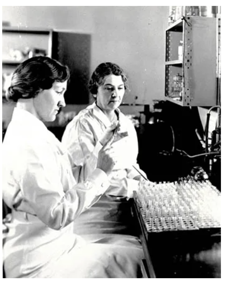
CREDIT: NATIONAL MUSEUM OF AMERICAN HISTORY
Margaret Pittman and fellow NIH physician Sadie L. Carlin are “reading” an agglutination reaction, part of the test for potency of commercially prepared anti-meningitis serum during the meningitis epidemic of 1935-1937 (1937).

As a woman in a male-dominated field, Margaret Pittman faced numerous challenges throughout her career. In an era when gender bias was pervasive, women struggled with standardisation and opportunities for advancement in scientific research. Pittman’s journey was the same, as she faced challenges that would have prevented her from moving further. The legacy of Dr. Margart Jane Pittman endures today, as her contributions continue to influence the direction of immunisation research. She stated that revolutionising problems with pertussis immunisation makes this work one of my best accomplishments that “despite the problems that have occurred with the pertussis vaccine… I consider this work one of my best accomplishments.”. Her partnerships and contributions have established a standard for upcoming generations and cleared the path for additional developments in vaccine research. By honouring her outstanding accomplishments, we can ensure that her influence on public health continues to be a driving force in the battle against infectious illnesses.

Written by Rabia Saleem