Towards 3D neuroblastoma cell models

It seems I have got a conference season. Three conferences within 2 months – no complains though. This time I went to the Matrix Biology Ireland Meeting in Galway. It was fantastic mix of topics and speakers ranging from new approaches in bone and heart repair to new matrixes in reconstruction of body tissues and diseases in the lab to minimise use of animals in pre-clinical studies.

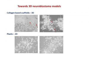
My talk was focused on neuroblastoma microenvironment and cell-to-cell communication through exosomes. I wrote about it in October post. I talked about things that did work and did not as well as new directions. One of the new directions is reconstructing neuroblastoma by growing neuroblastoma cells on collagen based scaffolds in 3D. Collagen constitutes most of our tissues to keep it shape and strength. These scaffolds are sponge-like matrixes built from collagen and other components. Of course cells grow differently on these matrixes. They have a different shape and growing properties in 3D. Neuroblatoma cells look like water drops on the cotton wool-like collagen scaffolds. In contrast, when they grow on plastic in 2D, they are flat. Studies show that cells in 3D respond to cytotoxic stress in a similar pattern as if being within a body (details in recent reviews 1-4).  It would be a great breakthrough once these models are optimised for neuroblstoma research  field. It will help to test all new and known drugs in the environment close to clinical settings. It could be a step forward to personilised therapies for children with neuroblastoma by isolating cancer cells, growing them in 3D and testing how they respond to all therapies available. It will facilitate more efficient design of treatment for relapsed or poorly responding tumours, sparing patients unnecessary rounds of chemotherapy and ultimately increasing survival.


Neuroblatoma cells look like water drops on the cotton wool like collagen scaffolds. In contrast, when they grow on plastic in 2D, they are flat. Arrows point towards cells.
This is microscopic images of neuroblatoma cells growing on the collagen scaffolds and plastic. Arrows point towards cells.


I’ve always felt that a selection of abstracts for an oral presentation is biased. The overall background and views of conference organisers would affect works selected for an oral presentation.  The same abstract was not selected for an oral presentation by one committee, but was supported by the other.  Never give up!

Readings:

  1. Schweiger PJ, Jensen KB.Modeling human disease using organotypic cultures. Curr Opin Cell Biol. 2016 43:22-29.
  2. Salamanna F, Contartese D, Maglio M, Fini M. A systematic review on in vitro 3D bone metastases models: A new horizon to recapitulate the native clinical scenario? Oncotarget. 2016 7(28):44803-44820.
  3. Picollet-D’hahan N, Dolega ME, Liguori L, Marquette C, Le Gac S, Gidrol X, Martin DK. A 3D Toolbox to Enhance Physiological Relevance of Human Tissue Models. Trends Biotechnol. 2016 34(9):757-69.
  4. Nyga A, Neves J, Stamati K, Loizidou M, Emberton M, Cheema U. The next level of 3D tumour models: immunocompetence. Drug Discov Today. 2016 21(9):1421-8.

How we work with cancer cell lines?

How we work with cancer cell lines?

The very first human cancer cell line was developed from a patient with an aggressive cervical cancer in 1951. This cell line was called HeLa after the patient name – Henrietta Lacks. This is the most popular and robust cancer cell line in biomedical research. Since then, other cancer cell lines were developed including neuroblastoma.

The first successful neuroblastoma ‘cell lines’ were cell populations from tumours that were adapted to grow for a short period in the lab environment in 1947. These tumour cell populations were used as a tool for diagnosis. This success inspired other researchers to develop long-term or immortal neuroblastoma cell lines. To date different neuroblastoma cell lines exist.

Cancer cell lines are sensitive and delicate in handling. They can only grow in the safe environment. Researchers have to protect them against bacteria, low temperatures, and too acidic/alkaline conditions. We protect cancer cells from bacteria contamination by handling them in cabinets where all plastic and media are sterile.

Handling neuroblastoma cells in the cell culture cabinet.

Cancer cells like to grow in conditions similar to conditions in human body. They like temperature of 36.6 – 37C. To achieve it special ‘green cell houses’ – CO2 incubators are built, which maintain the constant temperature, humidity and CO2 concentration.

We place cells in plastic dishes or containers called flasks and keep flasks in the ‘green cell houses’.

The cell growth and well being are checked regularly using microscopes. Healthy cells are to have similar shape, even distribution and grow attached to the plastic surface. Most microscopes have a camera attached to the top and linked to a computer. It helps to take picture of growing cells and record changes in cell behaviour.

Microscopic examination of drug resistant neuroblastoma cells KellyCis83. Cells look healthy and can be kept for another 2-3 days to form a more dense population.


Recommended reading

  1. Skloot, R. The Immortal Life of Henrietta Lacks 2011
  2. Thiele CJ. Neuroblastoma Cell Lines. Human Cell. 1998. 1-35 p.
  3. Murray M, Stout A. Distinctive Characteristics of the Sympathicoblastoma Cultivated in Vitro: A Method for Prompt Diagnosis. Am J Pathol. 1947;23(3):429–41.