Hi there, Ciara here again, a final-year PhD student in our research group. I can’t believe September has rolled around again, meaning one thing: it’s Childhood Cancer Awareness Month (CCAM). In honour of this month, I would like to tell you a little bit about the childhood cancer we study in our lab and the research that I do to one day help save children from this disease.
Neuroblastoma is an aggressive childhood cancer, with sadly only 20% of late-stage patients surviving after 5 years. Progressive disease and cancer relapse are common in neuroblastoma. This is due to standard treatment regimens not being adequate for treating high-risk patients. Current treatment also may cause a series of adverse reactions in patients. Therefore, my research focuses on developing a 3D model of high-risk neuroblastoma that models the cancer more accurately in a laboratory setting. This will act as a beneficial platform to test whether new therapies effectively fight the patients’ cancer cells, leading to better treatment options for children with neuroblastoma.
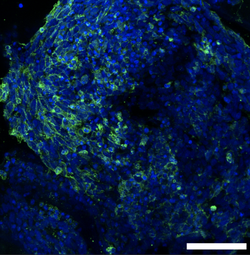
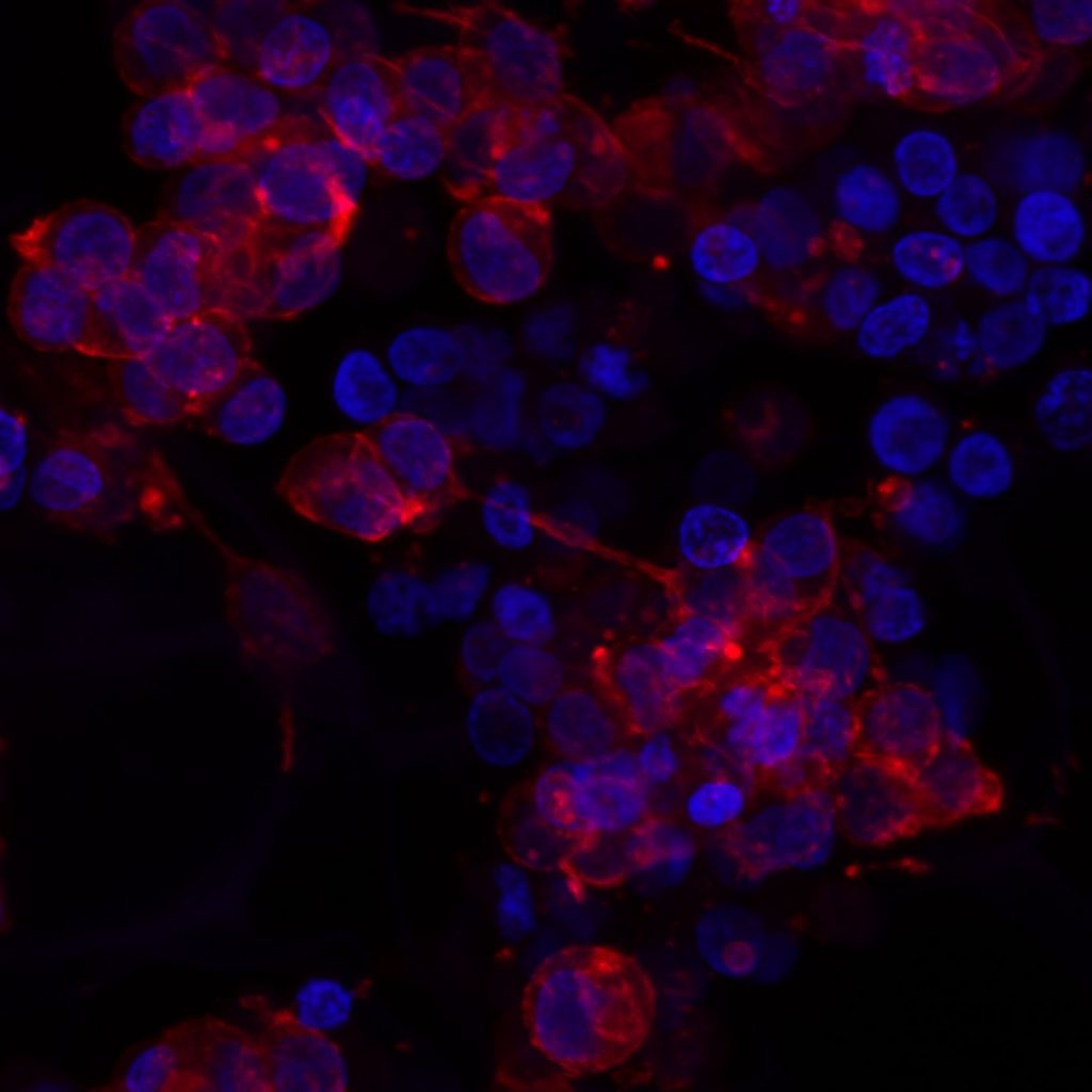
Below is a picture of how we grow these cancerous cells on our 3D model and visualise them with fluorescent stains. When we can see them like this under a microscope, we can study how they move and grow to help us understand how to treat them.
Here, we can see the cells growing on our 3D cancer model. This image is magnified by 200 times to be able to see the individual cancer cells. The green stain is the outside of our cancer cells, or we use the term, the cell membrane. The blue is the inside, or as some of you may know the term, the nucleus of the cell. (It is amazing what we can see with the power of microscopes, right?)
As you may know, every year, we support amazing charities by raising vital funds to keep the fight against childhood cancer going. Keep your eyes peeled on our Twitter for updates on what crazy activity we have committed to this year!!
Eight weeks ago, my journey into the intricate world of neuroblastoma began as I embarked on a remarkable research experience with the Cancer Bioengineering Group at RCSI. Guided by Dr. Olga Piskareva and supported by RCSI Research Summer School, this experience would transform my perspective on scientific exploration forever.
On my first day in the lab, excitement and nervousness mingled within me. But as I stepped into the bustling lab space, I was greeted with warm smiles and a sense of camaraderie among the researchers. The Cancer Bioengineering Group was known for its collaborative spirit, and it didn’t take long for me to feel like a valued member of the team.
RSS 2023 in Action
The research work was a perfect blend of diversity and fascination, encompassing both desk assignments and hands-on lab experiments. The highlight of it all was the chance to work with the cutting-edge 3D bio-printing machine, Rastrum. Witnessing the process of 3D bio-printing and using it to seed the Kelly cell line in various matrices left me in awe of the potential this technology held for future cancer therapies.
Yet, this journey extended beyond the realm of research. It was about the people – the passionate researchers who inspired and supported one another, the dedicated support staff who kept the lab running smoothly, and most notably, Dr. Olga Piskareva and Alysia Scott. They were more than mentors; they became friends and confidants, guiding me through challenges with unwavering support and celebrating our achievements as a team.
As the eight weeks drew to a close, I couldn’t help but reflect on the immense growth I had experienced professionally and personally. The cancer bioengineering field has unveiled the possibilities of using engineering principles to combat a disease that has touched countless lives worldwide.
This journey instilled in me a profound sense of purpose – a drive to contribute to the fight against neuroblastoma and other devastating illnesses. With a heart full of gratitude, I bid farewell to the Cancer Bioengineering Group at RCSI, knowing that the friendships forged and the knowledge gained would forever shape my future endeavours in the world of cancer research.
In the end, it wasn’t merely an eight-week stint; it was a transformational odyssey that solidified my passion for scientific discovery and my determination to make a difference in the lives of those affected by cancer. And for that, I will be eternally grateful.
Written by Mohammad Alabdulrahman, MED Class of 2026
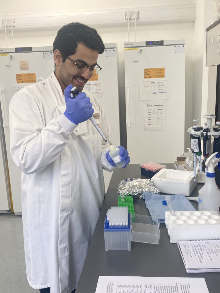
Hello everyone, I’m Amy! I joined the team for my TUD undergraduate research project in February, which is sadly coming to an end in the coming weeks. As my time here closes, I’m filled with mixed emotions. I am relieved and overjoyed to finish my thesis and see everything come together. However, I will certainly miss the team and working in the lab. I have learnt so much from my time here. For instance, research isn’t for the faint-hearted! It is filled with hiccups and bumps in the roads and unexpected twists and turns. This means you have to be able to make decisions and revise plans quickly. For that, I have so much respect for the whole team and anyone who chooses the path of research. I have also learnt so much about lab work and scientific writing. I was given independence throughout my work both in and out of the lab. With everyone more than willing to answer any queries I had and genuinely wanting to see me do my best.
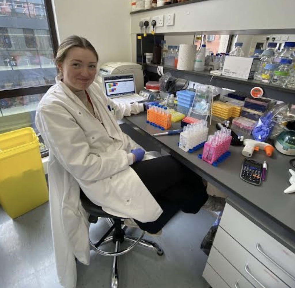
My favourite part of this research project has been the hands-on lab work, specifically the tissue culture. I’ve been trying to perfect my assay for DNA quantification recently. This photo was taken after I’d done tons of pipetting and got a hand cramp! My results looked nice, so it was all worth it. 🙂
Amy is at work!
All in all, I am very grateful for the opportunity to work with this amazing and dedicated team. I wish them all the best with their studies and research!
Finding suitable research models to study disease is a big challenge for researchers around the world. In cancer research, it is essential to work with models that can recapitulate tumour characteristics as much as possible. This is important to test chemotherapeutic drugs, understand tumour behaviour and have higher chances of translating the finds from the laboratory to clinical practice.
Multiple factors influence tumour behaviour and disease progression. The most important is the tumour microenvironment, which comprises different cells and molecules that surround the tumour and the extracellular matrix, a network of molecules that provides support to the cells in the body.
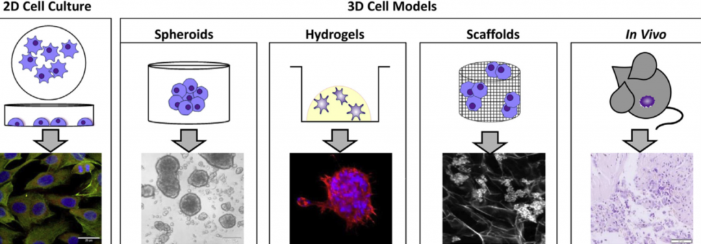
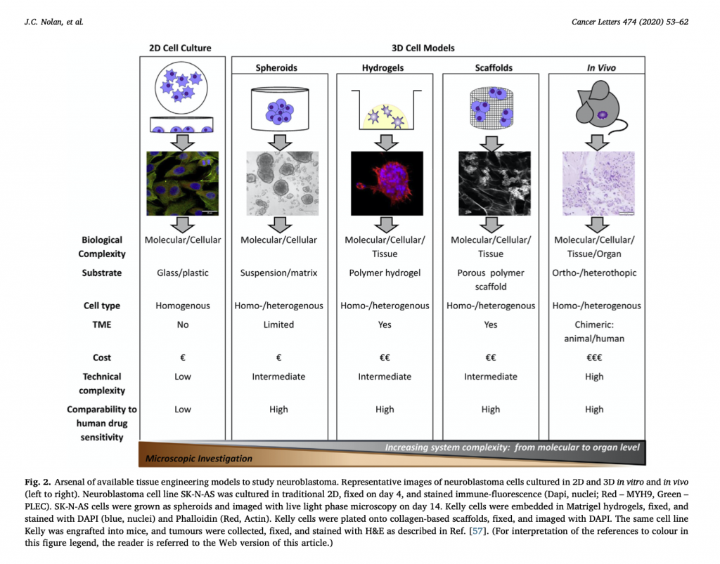
Most cell studies in a laboratory are based on 2D cell culture models in which the cells grow in a monolayer. Although this approach has a low cost and it is easy to use, it lacks the complexity observed in the clinical scenario. It is true that no model can recapitulate all the complexity found in the body. However, scientists were able to develop interesting approaches to study different tumour characteristics with relatively good approximation1.
Specifically for neuroblastoma, the most common solid tumour that affects children, scientists developed 3D models in which neuroblastoma cells grow interacting with the surrounding environment and with each other in a vial. Examples of 3D models include cells grown in hydrogels or scaffolds and multicellular tumour spheroids (see image below). Spheroids are formed through the self-adhesion of tumour cells growing in the form of very small balls. They can be maintained in the laboratory on their own or supported by scaffold-based platforms (jelly-like or porous materials). Scaffolds essentially support the cell resembling the extracellular matrix and surrounding tissue in the body.
Credits for the image: 3D models to study neuroblastoma. Adapted from Nolan, J. C. et al. Preclinical models for neuroblastoma: Advances and challenges. Cancer Lett. 474, 53–62 (2020).
In the Cancer Bioengineering Research Group, we work with neuroblastoma models such as organoids, a more complex type of spheroid, to understand neuroblastoma migration and invasion2. Moreover, we recently shared with the research community a protocol at jove.com describing the development of a 3D neuroblastoma model using collagen-based scaffolds3.
Time-lapse video of neuroblastoma organoids’ growth. Accompanying experimental data published in Gavin et al., Cancers 2021. Source: the Cancer Bioengineering Research Group
These models have the potential to advance drug tests performed in the laboratory providing better clinical translation, ultimately contributing to improving the quality of life and survival of children diagnosed with neuroblastoma.
The work with 3D models at the Cancer Bioengineering Research Group is supported by the Irish Research Council, the Conor Foley Neuroblastoma Cancer Research Foundation, Neuroblastoma UK and National Children’s Research Centre.
Written by Luiza Erthal
References
1. Nolan, J. C. et al. Preclinical models for neuroblastoma: Advances and challenges. Cancer Lett.474, 53–62 (2020).
2. Gavin, C. et al. Neuroblastoma Invasion Strategies Are Regulated by the Extracellular Matrix. Cancers13, 736 (2021).
3. Gallagher, C., Murphy, C., O’Brien, F. J. & Piskareva, O. Three-dimensional In Vitro Biomimetic Model of Neuroblastoma using Collagen-based Scaffolds. J. Vis. Exp. 62627 (2021) doi:10.3791/62627.
What a great start for 2020! Our long-lasting and productive collaboration with our colleagues from Tissue-Engineering Research Group Brough to live an important overview of the preclinical models for neuroblastoma. We particularly focused on the 3D in vitro models available.
During this exercise of searching and reading research papers, we found that researchers in neuroblastoma are looking for alternatives of traditional 2D culture. It is may be slow at the moment but the interest is there.
3D neuroblastoma models worked well in both validating known chemotherapies and screening new. The concepts and materials that were initially developed for bone or tissue regeneration can be used to a miniature model of neuroblastoma.
3D tissue-engineered models can accelerate drug discovery and development, reducing the use of animals in preclinical studies.
Full version is available at https://www.sciencedirect.com/science/article/pii/S0304383520300239?via%3Dihub
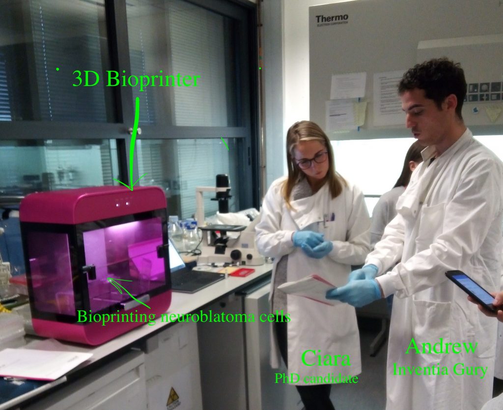
Here we go. Our first attempt to bio print neuroblastoma cells using Rastrum technology.
A compact pink oven-like device with a user-friendly interface and ‘magical’ disperse of cells and 3D environments. We bioprinted two types of neuroblastoma cells. One-easily forming clusters or tight groups and the other with high individualism in two types of homes: friendly and unfriendly. ‘Friendly’ homes have lots of clues to help cells to attach and grow. ‘Unfriendly’ homes have just a 3D niche aka house without furniture. Let’s see which homes cells like most.
An interesting idea or research question is always motivational. But it is a sketch till you get means to answer them. We, scientists, have to shape them into a proposal showing that we know limitations and have plans B & C if things go differently to planned. Then we apply for funding here and there… and many many times. The number of rejections makes us stronger – I hope. But one day, the idea may hit it right. So, it has happened to me recently and this SFI Award brings so needed fuel to study neuroblastoma.
The development and approval of new oncology drugs are very slow processes. This is mainly due to the big differences in the physiology of cancer cells grown on plastic and in the native microenvironment. Tissue engineering of tumour systems has a great potential to bridge this gap. This Award will help to advance our 3D tissue-engineered of neuroblastoma, that can be used in testing new drugs and new combinations of existing drugs.
Neuroblastoma cells grown in 3D
In particular, we will adapt the 3D model to screen different immunotherapies. This treatment option is very attractive both for adults and children because of its specificity and reduced side effects compared to chemotherapy, the current standard of care.
This Award will help my team to get a better understanding how neuroblastoma cells interact with the body environment, particularly with the immune system and how we can use the knowledge to develop new treatments and improve the patient outlook.
Reading my posts, it looks like I am more enjoying the cultural part and almost forgot the main reason I crossed the Atlantic with the Fulbright wings.
The first month in the lab was more a warming up. Where is my desk? Where is the cell culture rooms? How do they run it? How different is it? So, many microscopes – am I capable of imaging? And so on and so forth…
My typical day starts at 8-8.30 am and finishes once all is done. It may be 6pm or 10pm. Once the experiment is set up, I have to monitor cells every 24 hours for 5-7 days with no weekends or days off. The monitoring includes imaging. Lots of imaging. Every condition has 20-30 single cells to follow up. Each cell gets its own GPS tag manually to be able to image exactly the same cell as it grows and becomes a group of hundreds by multiplication. For example, I am running 8 different cell lines in 3 experimental conditions. So, 20-30 cells per all 24 combinations give us 480-720 individual cells to follow up. The imaging takes ~5 hours every day. After 5 days, I will have 2400 – 3600 pics of cells to analyse. It will be fun! I may need lots of Guinness to fly through that numbers.
Tagging cells. The left arrow points to a group of neuroblastoma cells. The arrow in the middle point to the same cells, but this image allows you to see the actual number of the cells. This group has 8 cells. The right arrow points to individual GPS tags for each cell
At the next step, I will select some of the conditions for video recording to trace cell fate from a single neuroblastoma cell to a metastatic niche consisting of hundreds of them. This video will show me how it all happens minute after minute.
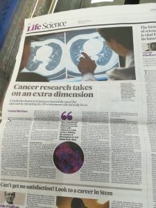
Appeared in today’s Irish Times. Lovely crafted by Dr. Vanesa Martinez
Although the discovery could be applicable in principle to any a solid tumour, Dr Piskareva’s target is neuroblastoma, a relatively common child cancer which affects a specific type of nerve cells in unborn children. “It’s quite aggressive and unfortunately there are many children who have metastasis when they are diagnosed, and this is the most challenging group to treat.”
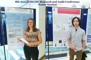
This was our 2nd time attending the OLCHC Research & Audit Day on May 25th, 2018. The conference provides a great forum for paediatric clinicians to share and update knowledge across different specialties through talks and poster presentations. It is insightful for basic biomedical researchers like us to see other perspectives.
I was delighted to know that two our studies were shortlisted. It is a rewarding feeling to see your Dream Team doing very well. One was the project of the Erasmus+ student Hanne Pappaert and the other was the project of NCRC funded Postdoc John Nolan. Hanne explored our 3D tissue-engineered model of neuroblastoma using collagen-based scaffolds with distinct mechanical properties. These new scaffolds were designed and manufactured by our collaborator Dr Cian O’Leary from Pharmacy Department and Tissue Engineering and Research Group (TERG) headed by Prof Fergal O’Brien. Hanne grew 5 neuroblastoma cell lines on the 3 scaffolds: hard like a rock, soft and fluffy like a cotton wool and a jelly-like. All cells liked the jelly-like environment. This environment is similar to bone marrow – the most common site of neuroblastoma metastasis. We were excited to see the difference as it means we are one step closer to reconstruct this type of tumour spread.
John has expanded our exploration of our 3D neuroblastoma model by examining the content of exosomes – little parcels sent by cancer cells in 3D and as tumours grown in mice. We were thrilled to see a high similarity in the exosomal content. This finding additionally proved the great applicability of our 3D model as a tool to study neuroblastoma.


 This was our 2nd time attending the OLCHC Research & Audit Day on May 25th, 2018. The conference provides a great forum for paediatric clinicians to share and update knowledge across different specialties through talks and poster presentations. It is insightful for basic biomedical researchers like us to see other perspectives.