Women in Science: Dr. Nina Marie Tandon

Throughout history, women in science have faced the hard challenge of navigating societal biases and limited opportunities in their pursuit of scientific discovery. Many of them were unjustly denied access to education and research positions just because of their gender, as it was thought that women were not capable of rigorous scientific work.

In the present, women in science (and in many other fields) still need to face many obstacles and challenges:

  1. Gender Bias: Women in science still encounter bias and stereotypes that can hinder their progress. They may face scepticism about their abilities and qualifications, leading to a lack of recognition for their work. Gender bias can also manifest in subtle ways, affecting opportunities for advancement and funding.
  2. Unequal Pay: Gender pay gaps persist in many scientific fields, with women often earning less than their male counterparts for similar work and qualifications.
  3. Limited Representation in Leadership Roles: Women remain underrepresented in leadership roles within academia, research institutions, and industry.
  4. Work-Life Balance: Balancing a career in science with family responsibilities can be particularly challenging for women. The demands of research, long working hours, and frequent travel can conflict with traditional gender roles and family expectations.

A woman and scientist who has certainly not let herself be stopped by all these adversities is Dr. Nina Marie Tandon. She has been a key player in the field of biomedical engineering, tissue engineering, and regenerative medicine. Her work focuses on developing innovative methods to grow artificial organs and tissues, using patient’s own cells to engineer tissues and organs. One of the cornerstones of Tandon’s work is the use of induced pluripotent stem cells. iPCs were developed in Japan pretty recently and are derived from skin or blood cells that have been reprogrammed back into an embryonic-like pluripotent state. This enables the development of an unlimited source of any type of human cell needed for therapeutic purposes.  

Tandon and her team employ cutting-edge bioreactors and 3D printing techniques to construct tissues: bioreactors provide a controlled environment for cells to grow and develop into specific tissue types, while 3D printing technology is used to create complex structures that mimic the architecture of natural organs.

In 2013, Tandon co-founded EpiBone, a biotech company that specializes in growing personalized bone grafts. EpiBone employs patient-specific stem cells to create skeletal structures based on individual DNA profiles, reducing the risk of rejection, streamlining surgical procedures, and potentially expediting patient recovery. They use a three-step process that begins with obtaining bone measurements and stem cells from abdominal fat via a CT scan, followed by creating a bone model in a bioreactor to stimulate growth. Finally, the patient’s own stem cells are added to the newly developed bone in the bioreactor, resulting in a fully functional replica bone ready for use.  Nina Tandon is also known for her engaging and informative TED Talks, where she discusses the future of medicine, tissue engineering, and the impact of regenerative medicine on healthcare.

I really admire Nina’s work, which I find genuinely fascinating. I believe her passion and determination come through in her TED talks, where she effectively manages to communicate complex scientific concepts and the potential of regenerative medicine to a broad audience.

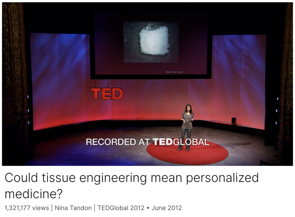
Click on the image to listen to one of her TED talks. Enjoy it 🙂

Written by Federico Cottone

Summer Research: A Journey of Insight with the Cancer Bioengineering Group

Eight weeks ago, my journey into the intricate world of neuroblastoma began as I embarked on a remarkable research experience with the Cancer Bioengineering Group at RCSI. Guided by Dr. Olga Piskareva and supported by RCSI Research Summer School, this experience would transform my perspective on scientific exploration forever.

On my first day in the lab, excitement and nervousness mingled within me. But as I stepped into the bustling lab space, I was greeted with warm smiles and a sense of camaraderie among the researchers. The Cancer Bioengineering Group was known for its collaborative spirit, and it didn’t take long for me to feel like a valued member of the team.

RSS 2023 in Action

The research work was a perfect blend of diversity and fascination, encompassing both desk assignments and hands-on lab experiments. The highlight of it all was the chance to work with the cutting-edge 3D bio-printing machine, Rastrum. Witnessing the process of 3D bio-printing and using it to seed the Kelly cell line in various matrices left me in awe of the potential this technology held for future cancer therapies.

Yet, this journey extended beyond the realm of research. It was about the people – the passionate researchers who inspired and supported one another, the dedicated support staff who kept the lab running smoothly, and most notably, Dr. Olga Piskareva and Alysia Scott. They were more than mentors; they became friends and confidants, guiding me through challenges with unwavering support and celebrating our achievements as a team.

As the eight weeks drew to a close, I couldn’t help but reflect on the immense growth I had experienced professionally and personally. The cancer bioengineering field has unveiled the possibilities of using engineering principles to combat a disease that has touched countless lives worldwide.

This journey instilled in me a profound sense of purpose – a drive to contribute to the fight against neuroblastoma and other devastating illnesses. With a heart full of gratitude, I bid farewell to the Cancer Bioengineering Group at RCSI, knowing that the friendships forged and the knowledge gained would forever shape my future endeavours in the world of cancer research.

In the end, it wasn’t merely an eight-week stint; it was a transformational odyssey that solidified my passion for scientific discovery and my determination to make a difference in the lives of those affected by cancer. And for that, I will be eternally grateful.

Written by Mohammad Alabdulrahman, MED Class of 2026

Preclinical models for neuroblastoma: Advances and challenges

What a great start for 2020! Our long-lasting and productive collaboration with our colleagues from Tissue-Engineering Research Group Brough to live an important overview of the preclinical models for neuroblastoma. We particularly focused on the 3D in vitro models available.

During this exercise of searching and reading research papers, we found that researchers in neuroblastoma are looking for alternatives of traditional 2D culture. It is may be slow at the moment but the interest is there.

3D neuroblastoma models worked well in both validating known chemotherapies and screening new. The concepts and materials that were initially developed for bone or tissue regeneration can be used to a miniature model of neuroblastoma.

3D tissue-engineered models can accelerate drug discovery and development, reducing the use of animals in preclinical studies.

Full version is available at https://www.sciencedirect.com/science/article/pii/S0304383520300239?via%3Dihub

8th OLCHC RESEARCH & AUDIT CONFERENCE

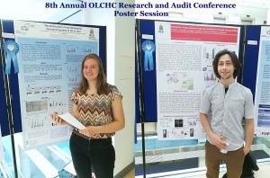
This was our 2nd time attending the OLCHC Research & Audit Day on May 25th, 2018. The conference provides a great forum for paediatric clinicians to share and update knowledge across different specialties through talks and poster presentations. It is insightful for basic biomedical researchers like us to see other perspectives.

I was delighted to know that two our studies were shortlisted. It is a rewarding feeling to see your Dream Team doing very well. One was the project of the Erasmus+ student Hanne Pappaert and the other was the project of NCRC funded Postdoc John Nolan. Hanne explored our 3D tissue-engineered model of neuroblastoma using collagen-based scaffolds with distinct mechanical properties. These new scaffolds were designed and manufactured by our collaborator Dr Cian O’Leary from Pharmacy Department and Tissue Engineering and Research Group (TERG) headed by Prof Fergal O’Brien. Hanne grew 5 neuroblastoma cell lines on the 3 scaffolds: hard like a rock, soft and fluffy like a cotton wool and a jelly-like. All cells liked the jelly-like environment. This environment is similar to bone marrow – the most common site of neuroblastoma metastasis. We were excited to see the difference as it means we are one step closer to reconstruct this type of tumour spread.

John has expanded our exploration of our 3D neuroblastoma model by examining the content of exosomes – little parcels sent by cancer cells in 3D and as tumours grown in mice.  We were thrilled to see a high similarity in the exosomal content. This finding additionally proved the great applicability of our 3D model as a tool to study neuroblastoma.


A new 3D strategy to study neuroblastoma

Our body has 3 dimensions: height, width and depth. Every single part of our body grows in the same 3 dimensions. This is true for cancer cells. Researchers use different ways to study cancer cells behaviour, how they grow and spread. We grow cells in the flasks, where they change their structure and shape and become flat losing one dimension. This is a very popular approach. We also grow cells in mice, where cells keep their 3D shape and mimic their behaviour to one observed in humans.

It is well known that we need to give a different amount of drug to kill cancer cells grown in flasks and in mice. This, in turn, delays the development of new drugs. Why does it happen this way? So, the drug works only on one side of the cell when they grow on the flat surface. In contrast, in mice, drug surrounds the cancer cell habitat and attacks cells at the edge first and then getting to those at the core. So we need more drug to kill cancer cells in mice.

We decided to design a new way to grow cancer cells that recreate their growth in 3 dimensions as in the human or mice body. We used special cotton wool like sponges as a new home for cancer cells and populated them with cancer cells. At the next step, we gave cells the drug at the different amount and checked what happened.

To understand cell fitness we stained them with red and blue dyes. On the left bottom side of the image, we see an equal amount of red and blue dyes telling us that cells were healthy and fit. Cells did not get any drug. When we gave a little amount of the drug but enough to kill cells in the flask, the balance of red and blue dyes was the same telling us that nothing really happened (the image in the middle). Cells were feeling well and healthy. The right bottom image has only blue dye. In this case, cells were given the amount of drug enough to destroy cancer cells in mice or humans. The lack of red dye tells us that this time the drug worked and killed the cancer cells.

We found that the drug killed cells on sponges only at doses enough to do the same in mice.

So, we concluded the new tactic to grow cancer cells in 3D on cotton-like sponges can bridge the gap between traditional way and animal models. This new strategy to grow cells on sponges should help to understand cancer cell behaviour better and accelerate the discovery and development of new effective drugs for neuroblastoma and other cancers. This, in turn, will make the outlook for little patients better and improve their quality of life.

This work has been published in Acta Biomaterialia and presented recently at the Oral Posters Session at the 54th Irish Association for Cancer Research Conference 2018.

This study was supported by Neuroblastoma UK and National Children’s Research Centre.

You can find more at

A physiologically relevant 3D collagen-based scaffold–neuroblastoma cell system exhibits chemosensitivity similar to orthotopic xenograft models.

IACR Meeting 2018 Programme

Neuroblastoma Research Dream Team 2017

It is fantastic to see so knowledgeable and enthusiastic young researchers in my research group. This year, the team is multinational with the Irish students mixing with Belgian and Malaysian. All together they are cracking the code of neuroblastoma microenvironment and tumour cells communication through understanding main differences between conventional cancer cell models and tumours.

The big research plan of the entire team consists of more smaller and focused projects to be completed within 10-12 weeks. All projects are unrestricted, they are driven by the intellectual curiosity of these students. This way is full of ups and downs, frustrations and encouragements when techniques do not work or reagents do not come in as expected. Some cancer concepts can also work differently in the given settings. Simple questions are bringing more challenges than expected.  But at the end of the road is the best reward – contribution to the conceptual advancement of neuroblastoma microenvironment.


The Neuroblastoma Research Dream Team 2017: Dr. John Nolan, NCRC funded researcher, RCSI, Joe O’Brien, TCD MSc student, Ciara Gallagher, DIT undergraduate student, Jessica Tate, RCSI Medical student, Larissa Deneweth, Erasmus student, Ghent, Ying Jie Tan, TCD MSc student.

Feeling good, excited and accomplished

This week can be rated for sure as feeling good, excited and accomplished. A UK based charity – Neuroblastoma UK has awarded a small grant to characterise a pre-clinical model of neuroblastoma which is a collaborative project between our lab and Tissue Engineering Research Group at RCSI. This project will study features of neuroblastoma cells growing on collagen-based scaffolds. The NBUK grant will contribute to one of the most expensive parts of the study – characterisation of cell secreting proteins using antibody-based profiling platforms.

Another research was accomplished yesterday –  John Nolan had his Voice Viva examination and successfully defended his PhD Thesis. This 3 year PhD project was funded by the National Children’s Research Centre. As his supervisor, I am delighted for him and wish him best of luck in his research career.