Sparkles of A Researcher Day

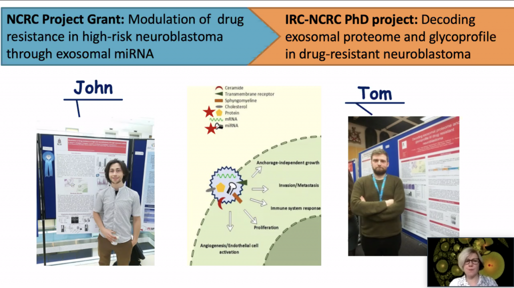
Once I mentioned the importance of the publication track record for a career in science. My team has been productive despite the COVID pandemic. Two review articles were published.

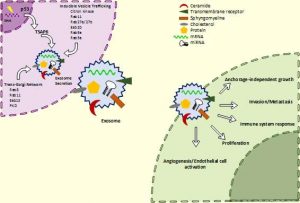
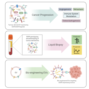
The first was a review written by Tom and published in Cancers focusing on the small extracellular vesicles produced by cancer cells that can transfer various growth signals to the tumour microenvironment aka neighbourhood and promote tumour expansion. The signal in focus was a protein called epidermal growth factor receptor (EGFR). It contributes to the healthy fitness of many different cells in the body. However, many cancer cells produce an excess of this protein giving them an advantage of growth over normal cells. Increased EGFR can be seen in breast, lung, glioblastoma and head and neck cancers.

Cancers 202012(11), 3200; https://doi.org/10.3390/cancers12113200

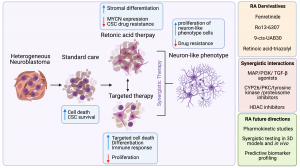
The second review has been published in Journal of Personalized Medicine on March, 16th. Originally, it was a small review project for Nadiya, a medicine student, last summer. However, it became a big one with all data systematically collected, analysed and condensed. The focus of this review was on Retinoic Acid (RA), widely known as Vitamin A and its role in neuroblastoma. RA plays a vital role in human development. The main feature of RA is to push neuroblastoma cells to become neuron-like cell stopping their aggressiveness and cancer fate. So, we wanted to know more about the ongoing research both in the labs and the clinic. We reviewed primary research articles reporting basic and translational findings as well as clinical trials. Hopefully, it would help other researchers to get a full picture of this topic and a structured resource of experimental models and drugs tested.

J. Pers. Med. 202111(3), 211; https://doi.org/10.3390/jpm11030211

Annual NCRC Symposia 2020

As the year comes to an end, you are looking back and seeing all achievements in a different light, a light of the COVID glaze. Lab research was at bay for a while, challenges to return and re-start experiments, no scientific meetings in the traditional format where you build your new collaborative net at coffee breaks. Despite all, the team has expanded and we welcomed Ellen and Erin in October.

The NCRC Winter Symposia is a lovely way to wrap the year putting together all hard work and look at the progress done so far. We have an exciting project that has two arms: a blue-sky science and a translational. Working together John and Tom were able to generate promising results on understanding how small membrane-bound vesicles or exosomes can send signals from neuroblastoma cells to cells responsible for new blood vessels formation. They developed a protocol to scale up the production of exosomes, isolate them and characterise. We have a dataset on what these exosomes carry on and now can test how they promote new blood vessels formation. Indeed, more left to do but knowing the direction makes this journey meaningful.

8th OLCHC RESEARCH & AUDIT CONFERENCE

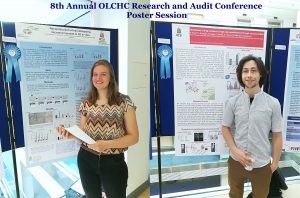
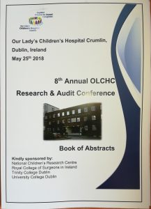
This was our 2nd time attending the OLCHC Research & Audit Day on May 25th, 2018. The conference provides a great forum for paediatric clinicians to share and update knowledge across different specialties through talks and poster presentations. It is insightful for basic biomedical researchers like us to see other perspectives.

I was delighted to know that two our studies were shortlisted. It is a rewarding feeling to see your Dream Team doing very well. One was the project of the Erasmus+ student Hanne Pappaert and the other was the project of NCRC funded Postdoc John Nolan. Hanne explored our 3D tissue-engineered model of neuroblastoma using collagen-based scaffolds with distinct mechanical properties. These new scaffolds were designed and manufactured by our collaborator Dr Cian O’Leary from Pharmacy Department and Tissue Engineering and Research Group (TERG) headed by Prof Fergal O’Brien. Hanne grew 5 neuroblastoma cell lines on the 3 scaffolds: hard like a rock, soft and fluffy like a cotton wool and a jelly-like. All cells liked the jelly-like environment. This environment is similar to bone marrow – the most common site of neuroblastoma metastasis. We were excited to see the difference as it means we are one step closer to reconstruct this type of tumour spread.

John has expanded our exploration of our 3D neuroblastoma model by examining the content of exosomes – little parcels sent by cancer cells in 3D and as tumours grown in mice.  We were thrilled to see a high similarity in the exosomal content. This finding additionally proved the great applicability of our 3D model as a tool to study neuroblastoma.


Congratulations to Dr.John Nolan!

My PhD student John Nolan together with other 41 candidates graduated at the RCSI’s 2017 June Conferring ceremony which took place in the College Hall of 123 St. Stephen’s Green.

He continues his research in neuroblastoma as a Postdoctoral researcher on the project funded by the National Children’s Research Centre. I am glad to be able to keep expertise and young talents in our team.

CMRF Spring Newsletter features neuroblastoma research

The research is a long-term investment. It is always built up on the work of the predecessors. Keep research running is crucial to make the dreams come true. Dreams for better treatment options and quality of life.


Thank you to everyone involved in raising funds for CMRF!

CMRF Spring Newsletter can be found here – CMRF-Spring Newsletter Final 15.05.17

Neuroblastoma Research Dream Team 2017

It is fantastic to see so knowledgeable and enthusiastic young researchers in my research group. This year, the team is multinational with the Irish students mixing with Belgian and Malaysian. All together they are cracking the code of neuroblastoma microenvironment and tumour cells communication through understanding main differences between conventional cancer cell models and tumours.

The big research plan of the entire team consists of more smaller and focused projects to be completed within 10-12 weeks. All projects are unrestricted, they are driven by the intellectual curiosity of these students. This way is full of ups and downs, frustrations and encouragements when techniques do not work or reagents do not come in as expected. Some cancer concepts can also work differently in the given settings. Simple questions are bringing more challenges than expected.  But at the end of the road is the best reward – contribution to the conceptual advancement of neuroblastoma microenvironment.


The Neuroblastoma Research Dream Team 2017: Dr. John Nolan, NCRC funded researcher, RCSI, Joe O’Brien, TCD MSc student, Ciara Gallagher, DIT undergraduate student, Jessica Tate, RCSI Medical student, Larissa Deneweth, Erasmus student, Ghent, Ying Jie Tan, TCD MSc student.

Identification of biomarkers of tumour response to drugs

A Research Project Grant funded by National Children’s Research Centre will be starting in April.

The ultimate aim is to identify biomarkers of tumour response to drugs in the blood of children with high-risk neuroblastoma.

Challenge: Treatment regimens for patients with high-risk neuroblastoma involve intensive, multi-modal chemotherapy. Many patients response to initial therapy very well, but has only short-term effects, with most becoming resistant to treatment and developing progressive disease.

The project has two parts which complement each other.

Part 1

  1. We will study cell-to-cell communication using cell-based models. We will collect exosomes, small envelopes containing bioactive molecules, produced by drug-resistant cell lines to treat non-cancerous cells. We will measure the effect of exosomes on non-cancerous cells by counting cell growth, examining their shape and metabolism. We will also examine whether non-cancerous cells treated with exosomes become less responsive to chemo drugs.
  2. We will treat neuroblastoma cells with a drug and collect exosomes before and after treatment. We will profile exosomes to identify any changes in their miRNA content. MiRNA are very small pieces of genetic material that can change the way cell feels and works. This step will help to find biologically active miRNA that can trigger cell resistance to drugs. These biologically active miRNA can represent biomarkers of tumour response to chemotherapy.

Part 2

  1. We will screen clinical samples for exosomal miRNA in response to drug treatment. We are planning to use a small sample of blood taken from neuroblastoma patients during routine examinations before, during and after chemotherapy.This step will help to find clinically relevant miRNA of tumour responsiveness to chemo drugs.

The plans for 3 years

How does this project contribute to the biomedical community?

This study aims to contribute to the better understanding of the disease mechanisms and scientific knowledge in the area, and in particular how neuroblastoma cells communicate with other cells helping tumour to create a unique microenvironment and protect themselves from chemotherapy pressure. The new data will give insights in biologically active proteins and miRNAs involved in cell-to-cell communication and drug responsiveness.

What are potential benefits of the proposed research to neuroblastoma patients?

This project aims to develop exosomal biomarkers of tumour response to drugs that might be used to help select patients for treatment and identify novel targets for the development of more effective personalised therapy with the anticipated improvement in outcomes. This work will contribute to the more efficient design of re-initiation treatment, sparing patients unnecessary rounds of chemotherapy and ultimately increasing survival. These new circulating markers will benefit children with high-risk neuroblastoma whose tumours are relapsed leading to less harmful and more tailored treatment options and improving their quality of life.

Cell to Cell Communicators

Tumour cells send different types of messages from one cell to another aka people post letters, postcards, and parcels to their families, friends, colleagues or  business. Cells can direct their messages using free moving proteins – postcards. They can wrap it in microvesicles with different cargo. Big microvesicles can take up big messages – parcels, small microvesicles or exosomes contain a limited number of texts – letters.

Tumour cells change their behaviour quickly adapting to anticancer therapies, so the messages they are sending. These messages can easily join blood stream and be read by researchers to understand how treatment is working and tumour cells are feeling.  Reading these messages from blood is more favourable as blood tests are done on the regular bases during and after the treatment.

In our lab we investigate how neuroblasts communicate with each other and the entire body through exosomes. We are interested to see what they write in their letters – exosomes. Do drug resistant and sensitive neuroblasts write different texts? What is the difference and how we can use this difference to predict child response to anticancer therapy?

In one set of experiments, we found that exosomes from drug resistant neuroblasts stimulate growth of sensitive cells. The more resistant neuroblasts send more powerful messages pushing cells to grow faster.

In the other set of experiments, we partially cracked the message showing that their texts are different. This finding explains why more resistant neuroblasts send more growth stimulating messages.

All these findings will be presented at the upcoming conference Goodbye Flat Biology: Models, Mechanisms and Microenvironment in Berlin.


schematic-exo2a

Schematic of exosome biogenesis and secretion. Cells produce exosomes through different pathways. This process is tightly regulated and controlled by numerous molecules. It can be triggered by many factors including extracellular stimuli (e.g., microbial attack, UV, drugs) and other stresses. The exosomes wrap up biologically active components such as proteins, RNA and miRNA. Exosomes can interact with recipient cells using four mechanisms: ligand/receptor interaction, protein transfer, membrane fusion or internalisation. Once exosomes entered the recipient cell, they release their content and re-programme the cell functions.


Suggested reading

Johnsen KB, Gudbergsson JM, Skov MN, Pilgaard L, Moos T, Duroux M. A comprehensive overview of exosomes as drug delivery vehicles – Endogenous nanocarriers for targeted cancer therapy. Biochim Biophys Acta – Rev Cancer. 2014;1846(1):75–87.

El Andaloussi S, Mäger I, Breakefield XO, Wood MJ a, Andaloussi S EL, Mäger I, et al. Extracellular vesicles: biology and emerging therapeutic opportunities. Nat Rev Drug Discov. 2013;12(5):347–57.

The schematic of exosomes was adapted from here