Research Summer School 2018

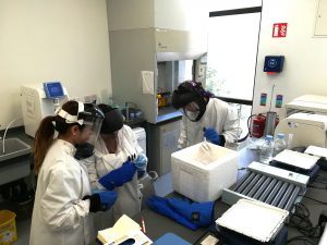
Another year, another Research Summer School students. We are hosting 4 students (Jessica, Dawn, Dola, and Jeff) this year. Some of them will be medical doctors, another will do research after the graduation. For them, the 8-weeks lab placement is a window into the reality of the everyday science. How cancer cells look? How do they grow? Where do we store them? How do we know that we have identified a new drug or a new target to study further? Do researchers have a sense of humour? Do they like donuts?

Why do they wear these astronaut helmets?

We have already said Good Bye to Jessica. Dola and Dawn’s projects are coming to an end this week, while Jeff is staying till the end of August.

Father of Chemotherapy and Cancer Immunology

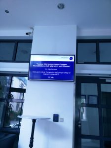
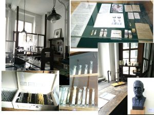
I was giving a talk at Georg-Speyer-Haus Institute for Tumour Biology and Experimental Therapy yesterday. The aim of my visit was to establish collaboration with Prof Daniela Krause, who is the expert in bone marrow microenvironment and targeted therapies. She took me to the Institute museum that keeps the history of this place and phenomenal researchers used to work there.

This research institute was established in 1904 to support work of Paul Ehrlich, its first director and funded by the private foundation “Chemotherapeutisches Forschungsinstitut Georg-Speyer-Haus”. Paul Erlich is the Father of the chemotherapy concept originally developed to treat diseases of bacterial origin. He reasoned that there should be a chemical compound that can specifically target bacteria and stop its growth. He developed Salvarsan, the most effective drug for treatment of syphilis until penicillin came onto the market.

Paul Erlich is also known for his contribution to cancer research. He and his colleagues actively experimented on how tumour originates and spread. They also tried to understand how immune system can beat cancer applying vaccination concepts.

Paul Erlich’s Lab back then. Now it is a museum

Paul Erlich and Ilya Mechnikov were jointly awarded The Nobel Prize in Physiology or Medicine for his “work on immunity” in 1908.


The Nobel Prize Diploma

Treatment of High-Risk Neuroblastoma

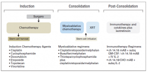
Children with high-risk neuroblastoma is the most challenging group to treat. Current treatment strategy for this group consists of 3 treatment blocks:

  1. induction: chemotherapy and primary tumour resection;
  2. consolidation: high-dose chemotherapy with autologous stem-cell rescue and external-beam radiotherapy [XRT];
  3. post-consolidation: anti–ganglioside 2 immunotherapy with cytokines and cis-retinoic acid.
Adopted from: Pinto NR et al JCO  2015, 33, 3008-3017.
Up to 50% of children that do respond experience disease recurrence with tumour resistant to multiple drugs and more aggressive behaviour that all too frequently results in death.
For the majority of children who do survive cancer, the battle is never over. Over 60% of long‐term childhood cancer survivors have a chronic illness as a consequence of the treatment; over 25% have a severe or life‐ threatening illness.
Reference:

Pinto NR, Applebaum MA, Volchenboum SL, Matthay KK, London WB, Ambros PF, Nakagawara A, Berthold F, Schleiermacher G, Park JR, Valteau-Couanet D, Pearson AD, Cohn SL. Advances in Risk Classification and Treatment Strategies for Neuroblastoma.J Clin Oncol. 2015 Sep 20;33(27):3008-17.


Drug resistant neuroblastoma cells

Children with neuroblastoma undergo several cycles of intensive chemotherapy to stop disease progression with the final aim to eliminate the tumour. Chemotherapy includes carboplatin or cisplatin in various combinations with drugs such as cyclophosphamide, ifosfamide, doxorubicin, etoposide, topotecan and vincristine (1). Nevertheless, in average 1 in 5 children with stage 4 disease do not respond to therapy. Up to 50% of children that do respond experience disease recurrence with tumour resistant to multiple drugs and more aggressive behaviour that all too frequently results in death.

The development of drug resistance is the major obstacle in treatment of neuroblastoma. To tackle this problem, researchers need to study different models of disease using cell lines, 3D tumour cell models, mice models and have access to clinical samples.

The first stage in testing drugs is to understand their killing ability of cancer cells. At this stage, researchers test drugs using cell lines. Cell lines are derived from tumours which were surgically removed from children with neuroblastoma. Researchers usually take a small piece of tumour straight after surgery and bring it into the laboratory.  Here, they place this piece into special solution that has enzymes to separate cells from each other. Then the suspension of all kind of tumour cells is placed into plastic dishes or flasks in a highly nutrient media to let cells grow. Cells that can adapt to these conditions start to grow, divide and produce a new generation of cancer cells. Researchers look after their growth, inspect their shape and behaviour; and test them on the presence of tumour markers. Once identity of these cells is confirmed they become a cell line and obtain a name. These cells keep majority of characteristics of the parental tumour and represent very useful tools in cancer research.

In our lab we use such cell lines to study neuroblastoma resistance to drugs. To understand changes in neuroblastoma biology during the development of drug resistance, we created drug resistant neuroblastoma cell lines (2). We treated three neuroblastoma cell lines CHP212, SK-N-AS and Kelly with cisplatin – a common drug in anticancer therapy. SK-N-AS and Kelly cells are sensitive to this drug, while CHP212 cells responded to this drug at much higher levels that the other two. Cells were grown in media containing cisplatin for several weeks. During this period most of the cells responded to cisplatin and died. Then we let cell survivors to recover in media without drug. This cycle was repeated several times until we got a population of cell survivors that can stand doses of cisplatin that can kill 50% of parental cells.  It took us more than 6 months to generate cisplatin resistant neuroblastoma cell lines CHP212Cis100, SK-N-ASCis24 and KellyCis83.

At the next step, we studied differences between these cell lines. We first compared their behaviour and cell shapes. Two resistant cell lines KellyCis83 and CHP212Cis100 started to grow faster, but SK-N-ASCis24 – slower than their parental cell lines. Interestingly, these cells also became more resistant to other drugs such as doxorubicin, etoposide, temozolomide, irinotecan and carmustin. These results are very important as they demonstrate that one drug can activate the cell defense systems that allow to escape toxicity of other drugs. These cell lines can be used to test new drugs and find those that can overcome developed resistance.

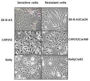
Cisplatin resistant cells also changed their appearance. Most dramatic changes occurred in SK-N-ASCis24 cells (see Figure 1).
nbl-cells

Figure 1. Microscopic images sensitive and drug resistant neuroblastoma cells (adapted from (2)) 

Two drug resistant cell lines SK-N-ASCis24 and CHP212Cis100 cells developed additional mobility skills – they became more invasive than their parental counterparts.
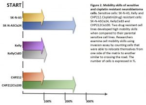
 

resistant-cells


Then we asked a question: what type of changes allowed cells to adapt to cytotoxic environment?  We examined changes in their genomic DNA first. We found that some genes increased their copy number, other went missing.

We identified changes in protein expression. More intriguingly, some proteins with the increased presence in the cells did not increase their presence in genomic DNA. We sorted these proteins on their role in cell processes such as migration, growth, cell cycle, etc. We found that each cisplatin resistant cell line developed a unique set of features that help them to escape cytotoxic stress (2). The similar patterns are found in clinic. Each patient responds to treatment differently.

What did we learn from this study?

  • One drug, in our study cisplatin, can activate the cell defense systems that allow to escape toxicity of other drugs.
  • The development of drug resistance gives cells new advantages and changes their behaviour and appearance, e.g. mobility skills, different cell shape, response to drugs, etc.
  • Each cisplatin resistant cell line developed a unique set of features that help them to escape cytotoxic stress.
  • These cell lines can be used to test new drugs and find those that can overcome developed resistance.

References

  1. Davidoff AM. Neuroblastoma. Semin Pediatr Surg. 2012; 21(1):2–14.
  2. Piskareva O, Harvey H, Nolan J, Conlon R, Alcock L, Buckley P, et al. The development of cisplatin resistance in neuroblastoma is accompanied by epithelial to mesenchymal transition in vitro. Cancer Lett. 2015;364(2):142–55. 


Cell to Cell Communicators

Tumour cells send different types of messages from one cell to another aka people post letters, postcards, and parcels to their families, friends, colleagues or  business. Cells can direct their messages using free moving proteins – postcards. They can wrap it in microvesicles with different cargo. Big microvesicles can take up big messages – parcels, small microvesicles or exosomes contain a limited number of texts – letters.

Tumour cells change their behaviour quickly adapting to anticancer therapies, so the messages they are sending. These messages can easily join blood stream and be read by researchers to understand how treatment is working and tumour cells are feeling.  Reading these messages from blood is more favourable as blood tests are done on the regular bases during and after the treatment.

In our lab we investigate how neuroblasts communicate with each other and the entire body through exosomes. We are interested to see what they write in their letters – exosomes. Do drug resistant and sensitive neuroblasts write different texts? What is the difference and how we can use this difference to predict child response to anticancer therapy?

In one set of experiments, we found that exosomes from drug resistant neuroblasts stimulate growth of sensitive cells. The more resistant neuroblasts send more powerful messages pushing cells to grow faster.

In the other set of experiments, we partially cracked the message showing that their texts are different. This finding explains why more resistant neuroblasts send more growth stimulating messages.

All these findings will be presented at the upcoming conference Goodbye Flat Biology: Models, Mechanisms and Microenvironment in Berlin.


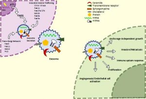
schematic-exo2a

Schematic of exosome biogenesis and secretion. Cells produce exosomes through different pathways. This process is tightly regulated and controlled by numerous molecules. It can be triggered by many factors including extracellular stimuli (e.g., microbial attack, UV, drugs) and other stresses. The exosomes wrap up biologically active components such as proteins, RNA and miRNA. Exosomes can interact with recipient cells using four mechanisms: ligand/receptor interaction, protein transfer, membrane fusion or internalisation. Once exosomes entered the recipient cell, they release their content and re-programme the cell functions.


Suggested reading

Johnsen KB, Gudbergsson JM, Skov MN, Pilgaard L, Moos T, Duroux M. A comprehensive overview of exosomes as drug delivery vehicles – Endogenous nanocarriers for targeted cancer therapy. Biochim Biophys Acta – Rev Cancer. 2014;1846(1):75–87.

El Andaloussi S, Mäger I, Breakefield XO, Wood MJ a, Andaloussi S EL, Mäger I, et al. Extracellular vesicles: biology and emerging therapeutic opportunities. Nat Rev Drug Discov. 2013;12(5):347–57.

The schematic of exosomes was adapted from here