Groundwork

Though the official announcement is scheduled for the first week of June, the groundwork is on. Lots of reading and planning for the trip to Johns Hopkins later this year. One of the first is the book by Rebecca Skloot ‘The Immortal Life Of  Henrietta Lacks”. The famous HeLa cells were generated by researchers at JH. The story is a fascinating journey for biomedical scientists and a tragedy for the Lacks family.

A new 3D strategy to study neuroblastoma

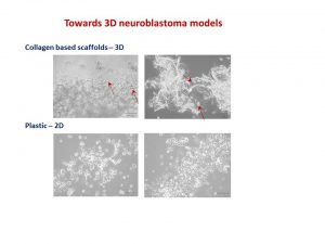
Our body has 3 dimensions: height, width and depth. Every single part of our body grows in the same 3 dimensions. This is true for cancer cells. Researchers use different ways to study cancer cells behaviour, how they grow and spread. We grow cells in the flasks, where they change their structure and shape and become flat losing one dimension. This is a very popular approach. We also grow cells in mice, where cells keep their 3D shape and mimic their behaviour to one observed in humans.

It is well known that we need to give a different amount of drug to kill cancer cells grown in flasks and in mice. This, in turn, delays the development of new drugs. Why does it happen this way? So, the drug works only on one side of the cell when they grow on the flat surface. In contrast, in mice, drug surrounds the cancer cell habitat and attacks cells at the edge first and then getting to those at the core. So we need more drug to kill cancer cells in mice.

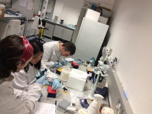
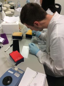
We decided to design a new way to grow cancer cells that recreate their growth in 3 dimensions as in the human or mice body. We used special cotton wool like sponges as a new home for cancer cells and populated them with cancer cells. At the next step, we gave cells the drug at the different amount and checked what happened.

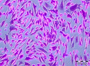
To understand cell fitness we stained them with red and blue dyes. On the left bottom side of the image, we see an equal amount of red and blue dyes telling us that cells were healthy and fit. Cells did not get any drug. When we gave a little amount of the drug but enough to kill cells in the flask, the balance of red and blue dyes was the same telling us that nothing really happened (the image in the middle). Cells were feeling well and healthy. The right bottom image has only blue dye. In this case, cells were given the amount of drug enough to destroy cancer cells in mice or humans. The lack of red dye tells us that this time the drug worked and killed the cancer cells.

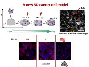
We found that the drug killed cells on sponges only at doses enough to do the same in mice.

So, we concluded the new tactic to grow cancer cells in 3D on cotton-like sponges can bridge the gap between traditional way and animal models. This new strategy to grow cells on sponges should help to understand cancer cell behaviour better and accelerate the discovery and development of new effective drugs for neuroblastoma and other cancers. This, in turn, will make the outlook for little patients better and improve their quality of life.

This work has been published in Acta Biomaterialia and presented recently at the Oral Posters Session at the 54th Irish Association for Cancer Research Conference 2018.

This study was supported by Neuroblastoma UK and National Children’s Research Centre.

You can find more at

A physiologically relevant 3D collagen-based scaffold–neuroblastoma cell system exhibits chemosensitivity similar to orthotopic xenograft models.

IACR Meeting 2018 Programme

Goodbye 2017! Hello 2018!

When I look back on my journey in 2017, there were many junctions, traffic lights and stops as well as ups and downs. Junctions were to make decisions, while traffic lights and stops – to be patient. Ups and downs were my feelings of satisfaction. The good mix of both kept me to stay human. It is not the number of grants received that matters it is who around you. I have met genuinely curiosity-driven students who made this journey fascinating and very special.

My most memorable Ups  were the successful examination and graduation of my PhD student John Nolan, organisation and chairing the IACR Meeting session: Challenges in Childhood Cancers, the Mad Hatter’s Tea Party and the Gala Dinner with the CFNCRF, the launch of my very own research team thanks to the funding by the NCRC and the Neuroblastoma UK, the successful completion of two final year undergraduate and two MSc projects, and welcoming the new PhD student Tom Frawley.

My team is growing and I am looking forward to 2018!

Goodbye 2017 and Hello 2018!

Treatment of High-Risk Neuroblastoma

Children with high-risk neuroblastoma is the most challenging group to treat. Current treatment strategy for this group consists of 3 treatment blocks:

  1. induction: chemotherapy and primary tumour resection;
  2. consolidation: high-dose chemotherapy with autologous stem-cell rescue and external-beam radiotherapy [XRT];
  3. post-consolidation: anti–ganglioside 2 immunotherapy with cytokines and cis-retinoic acid.
Adopted from: Pinto NR et al JCO  2015, 33, 3008-3017.
Up to 50% of children that do respond experience disease recurrence with tumour resistant to multiple drugs and more aggressive behaviour that all too frequently results in death.
For the majority of children who do survive cancer, the battle is never over. Over 60% of long‐term childhood cancer survivors have a chronic illness as a consequence of the treatment; over 25% have a severe or life‐ threatening illness.
Reference:

Pinto NR, Applebaum MA, Volchenboum SL, Matthay KK, London WB, Ambros PF, Nakagawara A, Berthold F, Schleiermacher G, Park JR, Valteau-Couanet D, Pearson AD, Cohn SL. Advances in Risk Classification and Treatment Strategies for Neuroblastoma.J Clin Oncol. 2015 Sep 20;33(27):3008-17.


August is a very quiet month

It is very quiet in the lab this month. No troubleshooting, no more long working hours, endless repetition of experiments, smiles and upsets… Almost all students completed their projects, submitted their works for grading and graduated. The last student is finishing at the end of August.

Time to focus on the collected data, reading literature, writing papers and new grants.

http://www.ifunny.com/pictures/its-rather-interesting-phenomenon-every-time-i/

Research Summer School in Action

It is always a pleasure to host undergraduate students during summer months. Two students joined the RCSI Research Summer School (RSS) Programme. Both are working on the NCRC funded project to understand mechanisms that drive neuroblastoma pathogenesis. None of them had a prior lab experience, but nothing is impossible under John’s supervision.

A full concentration on every single step of the research.

Why do we need fundraising for cancer research?

There is no short answer. Research is a slow, meticulous process of testing theories and finding out which ones work.It is exactly the same for both curiosity- and disease- driven questions. Long years of ground research full of ups and downs are critical for any breakthrough or progress. Very often with more downs than ups. Importantly, all researchers build on the work of their predecessors. This is the nature of science.

To understand the world around us, we have to do be curious and do “blue sky or curiosity-driven” research. It is a long shot, but this type of research can lead to practical applications down the road. One of the most recent examples is a drug Vismodegib (Erivedse) to treat basal cell carcinoma (the most common type of skin cancer) approved by the FDA in 2012. This drug targets genes of a hedgehog-associated signalling pathway. Defects in this pathway were found to drive many cases of skin cancer. But, how this relationship was found? Blue sky research!

Researchers studied hedgehog signalling in fruit flies and mice. One of the researchers had a strong interest in a fruit fly gene called hedgehog. If this gene is defective, then fly embryos look stubby and hairy aka a hedgehog. Further research brought more interesting facts and relationships leading to the identification of a drug that can stop the function of this faulty gene. Decades later with the advancement of genome sequencing, the defect in hedgehog signalling pathway genes was identified in patients with locally advanced and metastatic basal cell carcinoma.

What would happen if there were no research in fruit flies and mice? There would have been no rationale to create a drug like Vismodegib!

The best discovery research is unrestricted. It is driven by intellectual curiosity and conceptual advancement. More such curiosity- driven research is needed. For every medical breakthrough, for every Vismodegib, there were hundreds of blind alleys and failed ideas.

The research is a long-term investment. This contradicts to the short-term life of the politicians and governments who give the money. They do not take the risks. So, the discovery research becomes critically underfunded.

Fundraising creates opportunities for blue sky research and developing cancer treatments.

Thank you all who support cancer research charities!


Feeling good, excited and accomplished

This week can be rated for sure as feeling good, excited and accomplished. A UK based charity – Neuroblastoma UK has awarded a small grant to characterise a pre-clinical model of neuroblastoma which is a collaborative project between our lab and Tissue Engineering Research Group at RCSI. This project will study features of neuroblastoma cells growing on collagen-based scaffolds. The NBUK grant will contribute to one of the most expensive parts of the study – characterisation of cell secreting proteins using antibody-based profiling platforms.

Another research was accomplished yesterday –  John Nolan had his Voice Viva examination and successfully defended his PhD Thesis. This 3 year PhD project was funded by the National Children’s Research Centre. As his supervisor, I am delighted for him and wish him best of luck in his research career.


May whatever we do at the lab today make a difference in another person’s life someday in the future.

Mei Rin Liew

I am a medical student at Penang Medical College under a twinning programme with the Royal College of Surgeons in Ireland. I studied my pre-clinical years at RCSI Dublin. In the summer of 2015, I had the opportunity to join the RCSI Research Summer School (RSS) Programme. I was mentored by Dr Olga Piskareva, from Cancer Genetics, Molecular and Cellular Therapeutics (MCT) Department, RCSI.  Being in this lab was simply one of the greatest experiences I have in my life; it was really rewarding.

My RSS project investigated the role of VDAC-1 protein on chemotherapy resistance in neuroblastoma. The only research focus of this lab is to find key players in neuroblastoma pathogenesis and to advance anti-cancer therapy.

Neuroblastoma cell line SK-N-AS. The cell line in my experiments.

I was entrusted with the task of splitting cells. I would plate them onto 96-well plates, add cisplatin drug and measure their viability afterwards. It may sound simple here, but the whole process required passion and hard-work.

Prior to this, I did not have any experience in the medical research field. During my first two weeks, everything seemed so tough; however, they became easier as the weeks flew by. My mentor, Olga, and the other staff and PhD students (Garret, John and Ross) were helpful and always guided me to explore my potentials. This programme taught me various new things which I would not have acquired on a normal day-to-day basis in school.

Introduction to Malaysian cuisine.

The people at Cancer Genetics were warm and wonderful. The hospitality, love and guidance cannot be quantified and words cannot express my immense gratitude towards them. It has been fascinating and I cherish every moment I spent there. We bonded over our weekly breakfast and tea sessions so well, and I am indeed grateful for being a part of this big family. It is my sincere wish that this positive spirit of togetherness will be preserved and will grow stronger in the future. This is something special, and I think ours is the best lab at RCSI!

Under this RSS, all the participants attended skills workshops and weekly Discovery Series lectures. We were also given a Biography of Cancer by Siddhartha Mukherjee to read; evidently a good read. Here are the links to the RCSI Research Summer School Student Testimonial Videos.

I returned to Penang Medical College to further my studies in my clinical years. I took part in the PMC Research Day 2016 in which I was awarded the First Prize in Oral Presentation. I would like to dedicate this success to Olga and everyone who has been with me throughout my time at Cancer Genetics. Without all the guidance, I would not have made it this far.

I strongly urge students to take part in the research opportunities, because you gain invaluable experiences that you do not get elsewhere. May whatever we do at the lab today make a difference in another person’s life someday in the future.

Mei Rin Liew

Towards 3D neuroblastoma cell models

It seems I have got a conference season. Three conferences within 2 months – no complains though. This time I went to the Matrix Biology Ireland Meeting in Galway. It was fantastic mix of topics and speakers ranging from new approaches in bone and heart repair to new matrixes in reconstruction of body tissues and diseases in the lab to minimise use of animals in pre-clinical studies.

My talk was focused on neuroblastoma microenvironment and cell-to-cell communication through exosomes. I wrote about it in October post. I talked about things that did work and did not as well as new directions. One of the new directions is reconstructing neuroblastoma by growing neuroblastoma cells on collagen based scaffolds in 3D. Collagen constitutes most of our tissues to keep it shape and strength. These scaffolds are sponge-like matrixes built from collagen and other components. Of course cells grow differently on these matrixes. They have a different shape and growing properties in 3D. Neuroblatoma cells look like water drops on the cotton wool-like collagen scaffolds. In contrast, when they grow on plastic in 2D, they are flat. Studies show that cells in 3D respond to cytotoxic stress in a similar pattern as if being within a body (details in recent reviews 1-4).  It would be a great breakthrough once these models are optimised for neuroblstoma research  field. It will help to test all new and known drugs in the environment close to clinical settings. It could be a step forward to personilised therapies for children with neuroblastoma by isolating cancer cells, growing them in 3D and testing how they respond to all therapies available. It will facilitate more efficient design of treatment for relapsed or poorly responding tumours, sparing patients unnecessary rounds of chemotherapy and ultimately increasing survival.


Neuroblatoma cells look like water drops on the cotton wool like collagen scaffolds. In contrast, when they grow on plastic in 2D, they are flat. Arrows point towards cells.
This is microscopic images of neuroblatoma cells growing on the collagen scaffolds and plastic. Arrows point towards cells.


I’ve always felt that a selection of abstracts for an oral presentation is biased. The overall background and views of conference organisers would affect works selected for an oral presentation.  The same abstract was not selected for an oral presentation by one committee, but was supported by the other.  Never give up!

Readings:

  1. Schweiger PJ, Jensen KB.Modeling human disease using organotypic cultures. Curr Opin Cell Biol. 2016 43:22-29.
  2. Salamanna F, Contartese D, Maglio M, Fini M. A systematic review on in vitro 3D bone metastases models: A new horizon to recapitulate the native clinical scenario? Oncotarget. 2016 7(28):44803-44820.
  3. Picollet-D’hahan N, Dolega ME, Liguori L, Marquette C, Le Gac S, Gidrol X, Martin DK. A 3D Toolbox to Enhance Physiological Relevance of Human Tissue Models. Trends Biotechnol. 2016 34(9):757-69.
  4. Nyga A, Neves J, Stamati K, Loizidou M, Emberton M, Cheema U. The next level of 3D tumour models: immunocompetence. Drug Discov Today. 2016 21(9):1421-8.