’m writing to you from the Biomedicum, a research facility at the University of Helsinki. As you can probably guess, I didn’t travel here for the Finnish weather, but to collaborate with a team that is part of the European Mac4Me consortium. This group specialises in a fascinating imaging technique called multiplex immunohistochemistry. In simple terms, they take patient tissue samples or samples created in the lab and cut them into incredibly thin slices—just 4 micrometres thick, which is about 4/100ths of a millimetre. They then use multiple rounds of staining with antibodies to visualise different tumour markers and map out exactly where these markers are located within the sample. You can see an example of this stained tissue below.
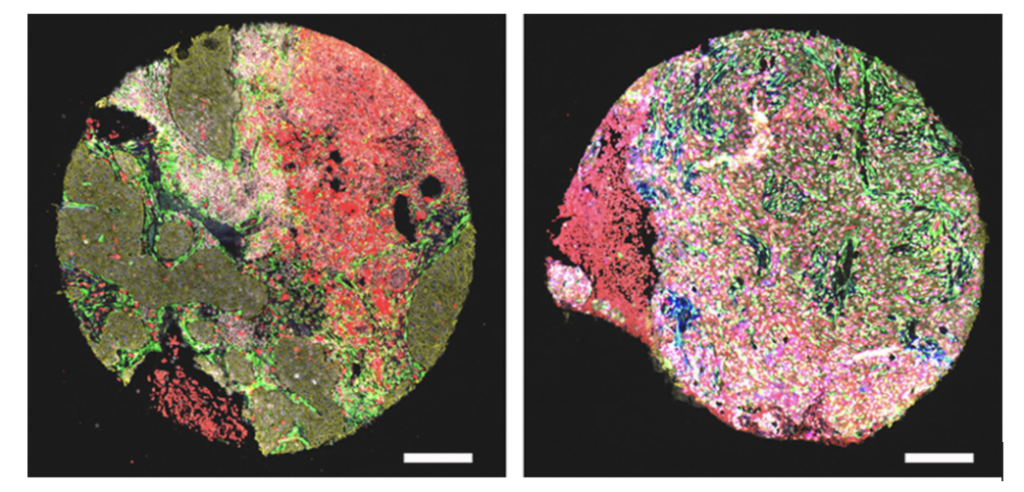
Viiklepp K Journal of Investigative Dermatology. 2025
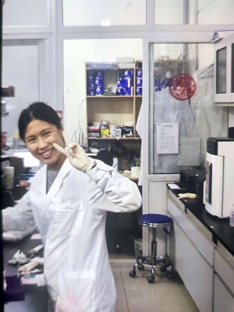
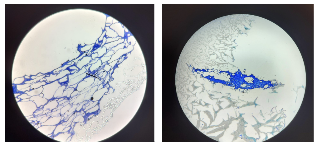
For this project, I brought along different versions of a “brain-like” hydrogel I’ve created in the lab. These hydrogels have varying stiffness and molecular compositions, and they contain Neuroblastoma tumour cells growing within them (you can see simple cell staining below). My goal here is to use the lab’s advanced imaging machine to observe how these tumour cells behave. I want to see if they are multiplying, if they seem stressed or healthy, and how they adapt to the different molecular environments of the hydrogels.
This approach will be incredibly valuable for studying how the tumour’s surroundings—the microenvironment—influence the spread of Neuroblastoma to the brain. It will also help us understand what happens when we add new molecular components to the hydrogel to more closely mimic the brain environment. Looking ahead, I plan to use this same technique for an even more complex experiment: by growing immune cells together with the neuroblastoma cells, I hope to visualise and identify the specific pathways the cancer cells use to communicate with and potentially suppress the immune system.
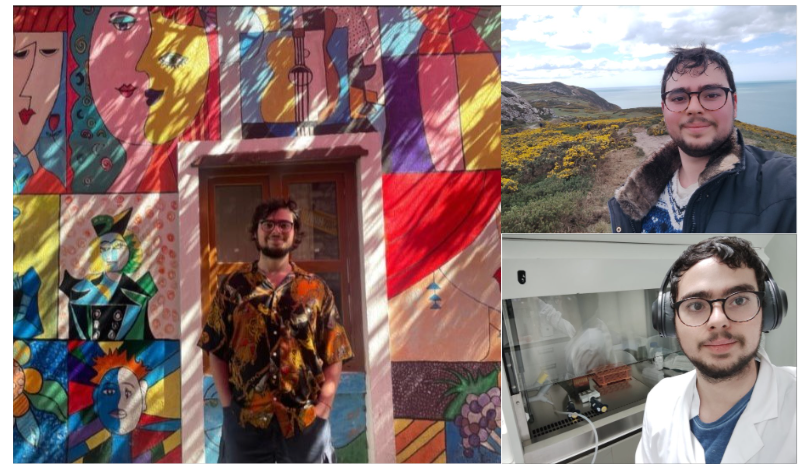
Written by Pierluca Cancellieri, Mac4Me MSCA PhD