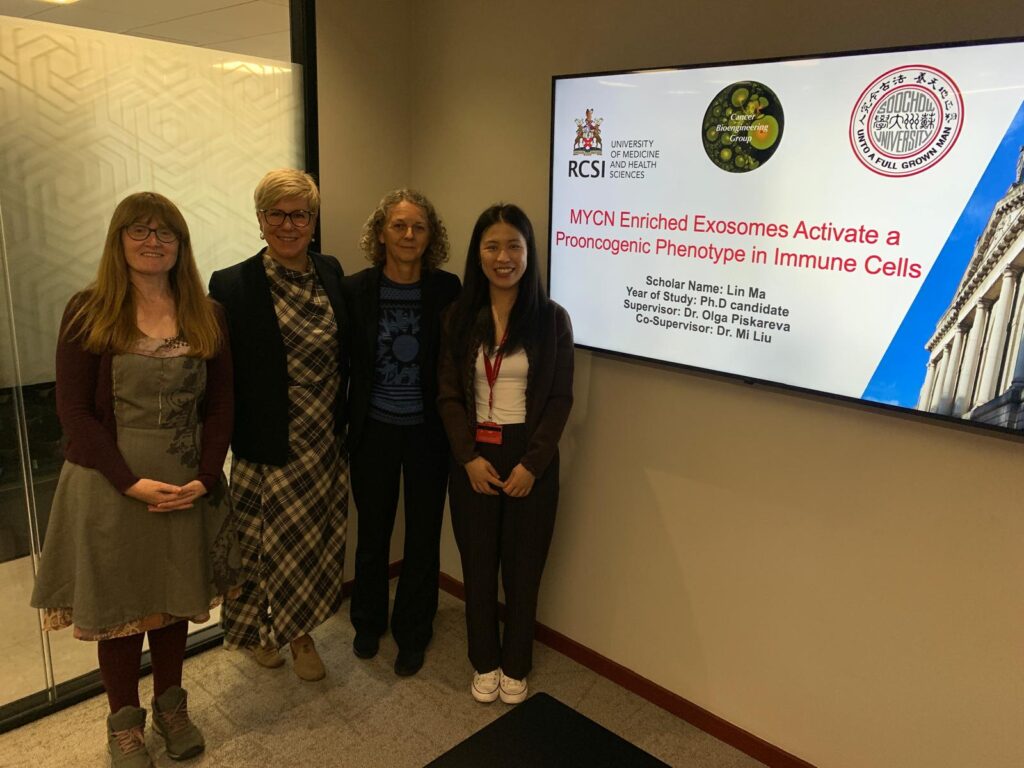
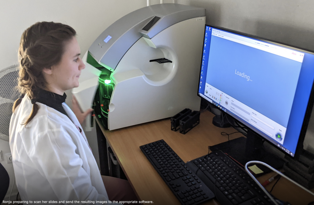
What a year – two young and talented postgraduate students have been minted with a Doctor of Philosophy Degree in September and December of 2025. They are Dr Lin Ma and Dr Ronja Struck. Hard work and dedication are the cornerstones of this challenging but rewarding journey.
They sailed through scattered showers and sunny spells, gale winds and stormy snow with sunshine developing elsewhere, turning chilly under clear skies on some days with temperatures below/above zero. The full spectrum of emotions and hard work was spiced up by the uncertainty of COVID-19 restrictions. Well done to Ronja and Lin!
Hot off the press! The study carried out by Thomas Frawley during his PhD has just been published in Journal of Personalized Medicine.
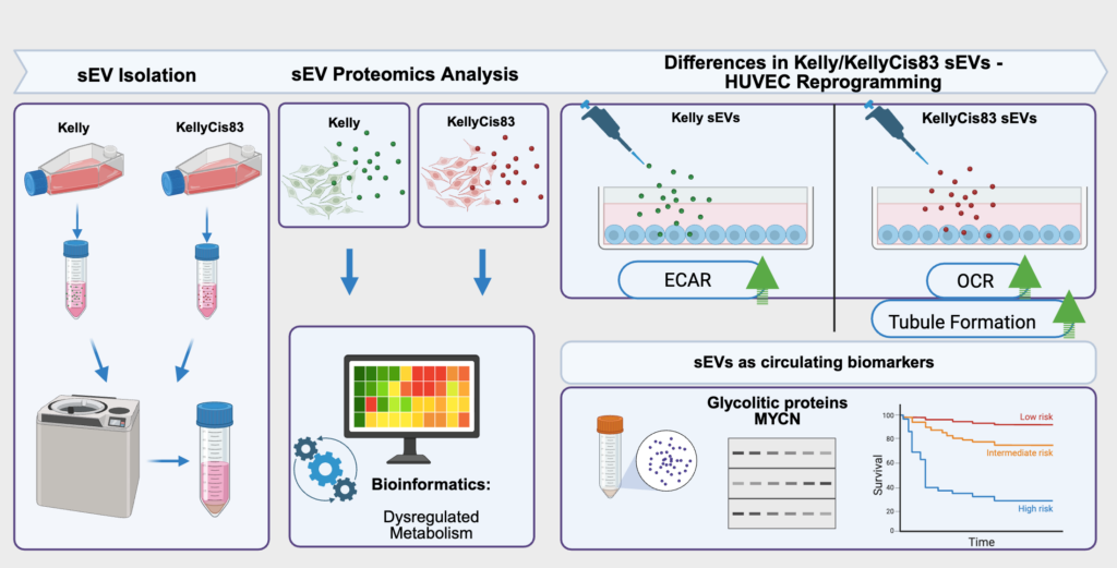
Cancer that is resistant to treatment is a big challenge because it often leads to lower survival rates. Tumour cells release small extracellular vesicles, which can influence other cells in the body by carrying various proteins. The study focused on understanding what proteins are in these particles from resistant and sensitive cancer cells and how they affect non-cancerous cells, like those involved in forming blood vessels. Our study discovered that these packages from resistant cancer cells contain special proteins involved in how cells produce and use energy. These findings suggest that these proteins could be used as markers to monitor disease progression or treatment response, using less invasive methods such as blood tests.
A schematic summary of Frawley’s study, also known as a graphical abstract. J. Pers. Med. 2025, 15(12), 584; https://doi.org/10.3390/jpm15120584 (registering DOI)
Understanding how resistant cancer cells influence their surroundings could lead to new ways of diagnosing and treating high-risk neuroblastoma. Detecting these proteins through blood tests could help personalise treatment strategies, making them more effective without the need for invasive procedures. This research opens the door to using tiny particles from blood to better understand how cancer progresses and responds to therapy.
Good afternoon, readers! Pierluca here, writing to you as one of the newest members of this incredible team. For those who haven’t met me yet, I’m a PhD student joining the RCSI family for the next three years.
My story starts in Brindisi, a charming harbor town in southeastern Italy. From there, my academic journey took me to the Netherlands. During my two research projects, I explored how high-fat diets impact liver metabolism and investigated ways to prevent metabolic reprogramming and cell death.
Now, I’m bringing that curiosity to RCSI, where my focus is shifting to something even more complex: cancer metastasis. In the lab, I’ll research how neuroblastoma invades the brain to form metastases. Using 3D bioprinting and scaffold models, I’ll grow Neuroblastoma Kelly and Kelly-cis cells to observe how they infiltrate brain-like structures and hijack the immune system.
Science is intense, so balance is key! When I’m not in the lab, you’ll find me Hiking when the sun is shining or Playing cards in a cozy pub when the rain pours. Cooking with friends is a great way to spend some relaxing time at home and when I am alone, I enjoy a good book (currently reading The Master and Margarita, highly recommend!).
Stay tuned for more about me and my research!
Written by Pierluca Cancellieri, Mac4Me PhD student
Hi everyone! I’m Chunyu, and I’ve recently started my PhD journey in the field of bioengineering and neurobiology. My academic background includes an MRes in Biomedical Research from Imperial College London, where I developed a deep interest in microfluidic technologies and their applications in disease modelling.
Currently, my PhD project focuses on identifying the function of macrophages—the body’s frontline immune cells—when they first interact with neuroblastoma (NB) cells using a brain and liver organ-on-a-chip (OoC) model. By recreating these organ environments on a chip, I aim to explore how macrophages respond to NB invasion and how this early interaction might shape the progression of the disease. This research could open new doors for early intervention and treatment strategies in childhood cancers like neuroblastoma.
When I’m not in the lab, you’ll probably find me outside—going on hikes, enjoying a good swim, or finding a tasty Hotpot restaurant. I love blending my curiosity for science with a love for the outdoors, and I’m excited to share updates from both worlds as I go through this PhD journey.
Thanks for stopping by, and stay tuned for more science and a few outdoor adventures along the way!
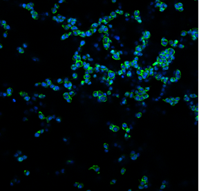
Sometimes, the most fascinating parts of science are invisible to the naked eye—like in these images captured with a confocal microscope!
What you’re seeing here are DC 2.4 cells, a mouse dendritic cell line. These immune cells are key players in recognising foreign substances (like bacteria, viruses, or even cancer cells) and activating the body’s immune response.
In this experiment, we cultured the DC 2.4 cells on a sponge-like material composed of collagen and glycosaminoglycans (GAG), two natural components commonly found in body tissues. This material is called a scaffold, and it provides cells with a 3D surface to grow on, more closely mimicking their natural environment within the body.
To make the cells visible under the microscope, we used two fluorescent stains:
DAPI (blue), which marks the nucleus—the control centre of the cell,
Phalloidin (green), which highlights the actin filaments that give the cell shape and structure.
We’re testing how well these immune cells survive, attach, and spread on the collagen-GAG scaffold over time. By utilising a 3D environment, we can gain a deeper understanding of how cells behave in more realistic conditions. This is especially important for research into cancer immunotherapy and vaccine development.
This image tells us that the DC 2.4 cells can successfully grow and interact with the scaffold!
As a DevelopMed Marie Skłodowska-Curie Fellow, I am committed to advancing childhood cancer research by investigating the biology of neuroblastoma, a complex and aggressive paediatric solid tumour. My research focuses on the high-risk form of the disease, where amplification of the MYCN oncogene is strongly associated with poor prognosis.
The project aims to elucidate the pathway crosstalk regulated by MYCN—specifically, how it alters normal cellular signalling and governs the critical cell fate decisions between proliferation and apoptosis. By employing mass spectrometry-based proteomics combined with systems biology approaches, I am constructing a comprehensive map of MYCN-driven signalling networks to identify potential therapeutic targets that could improve clinical outcomes for affected children.
A distinctive and rewarding aspect of my fellowship is my role as a visiting scientist at the Royal College of Surgeons in Ireland (RCSI), where I collaborate with Dr. Olga Piskareva’s lab, an internationally recognised leader in 3D neuroblastoma research. Here, I am gaining hands-on experience with 3D neuroblastoma spheroid culture systems, which more accurately recapitulate tumour behaviour compared to traditional 2D models. These advanced systems enable a deeper understanding of drug responses, tumour architecture, and cellular interactions in a physiologically relevant context.
This collaborative framework between UCD and RCSI fosters a dynamic, translational research environment and exemplifies the core values of the Marie Curie programme—innovation, collaboration, and real-world impact.
Every stage of this journey—from pathway elucidation to 3D model validation—contributes to the overarching goal of developing more effective, targeted therapies for children diagnosed with neuroblastoma.
The European Association for Cancer Research (EACR) is a registered charity and scientific community that has been holding conferences since 1968. EACR’s annual four-day congress is dedicated to basic, preclinical and translational cancer research. It brings together the cancer research community, including PhD students, postdocs, PIs, and commercial sponsors, for the opportunity to network and collaborate to progress cancer therapeutics.
I was fortunate enough to receive the Breakthrough Cancer Research Education and Travel Award, which made it possible for me to attend this year’s EACR conference held in Lisbon, Portugal. Breakthrough Cancer Research is an Irish Medical research charity focused on improving the outcomes of patients diagnosed with rare and poor prognosis cancers, like neuroblastoma.
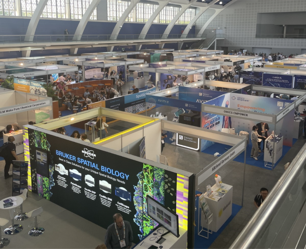
When I first arrived at the congress center in Lisbon, I was immediately impressed by how well organized and put together the conference was. A schedule of four full days included speakers, poster presentations, industry talks, a technology exhibition, giveaways, networking rounds, and early-career talks. I checked in, received my “goodie bag” and was on my way to the first talk. For the duration of the conference, you were encouraged to move freely between all the available presentations within several auditoriums and pavilions. They even had screens and speakers set up outside the auditoriums if there was no more space inside to make sure that the research presented was accessible to everyone. The lunch breaks were the perfect time to enjoy the sunshine, walk along the Tagus River, and have a picnic with views of the Ponte 25 de Abril bridge (similar in style to the Golden Gate Bridge in San Francisco, California).
Exhibitors showcase with over 100 companies available to talk about their technology. QR codes were at each booth to scan for participants to be entered into a drawing for an iPad and free entry to next year’s conference in Budapest, Hungary.
Throughout the conference, I listened to talks that ranged from how estrogen levels in breast cancer are related to the loss of bone density to how we can detect cancer in circulating cells for a diagnosis three years earlier than previous tests. One of the talks began with the necessity for physiologically relevant in vitro to 3D models and then the conclusion of the talk discussed how there’s a bridge needed between academia and industry for treatments to be more streamlined and accessible. Most importantly, I was able to read quite a few posters with research that other PhD students were doing related to small extracellular vesicles (sEVs). My work specifically looks at the relationship between sEVs shared from cancerous to non-cancerous cells and what their functional impact is. A lot of the work I saw was optimization of sEV isolation and characterization, which can be quite tricky to do but was helpful to see what complications others were running into and their troubleshooting results.
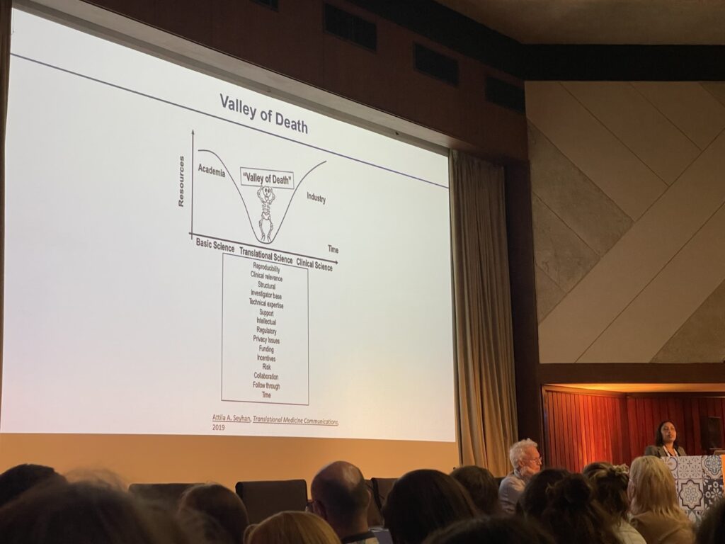
Presentation by Cindrilla Chumduri during EACR – EMBO Symposium: Advanced in vitro Models. Chumduri highlights the “valley of death” where there is a gap between academic and industry research that impedes the progression of scientific breakthroughs in cancer research.
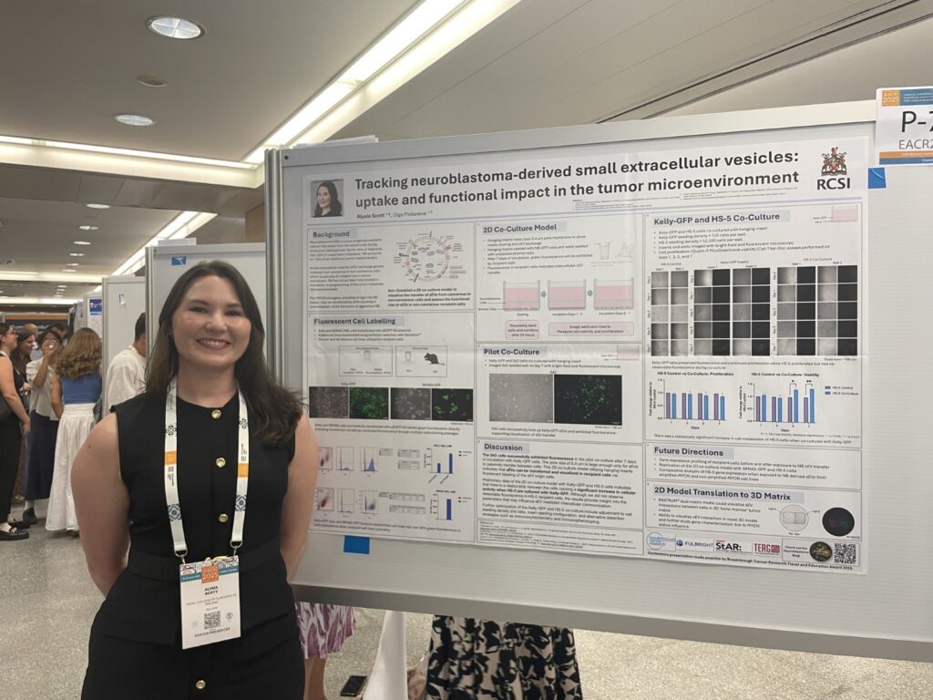
By the time it got to my poster defense, I was excited to talk about my work and looking forward to meeting others who might be doing research similar to mine. There were a handful of people that came to speak to me about my work and ask questions. One thing about the PhD journey is that sometimes you can be so deeply involved in your own work and what isn’t going right that you lose sight of how impactful your work can be. When several people approached me about the co-culture model I was using, they were so curious and wanted to implement something like that into their work. Hearing positive feedback on my efforts was a refreshing way to end the conference. At the end of the day, there was a celebration dinner where a traditional Portuguese Fado band played music while we were able to unwind and network with other PhD students. My time spent in Lisbon at EACR was one of the best conference experiences I’ve had. I’m looking forward to heading back into the lab, making progress with my project, and presenting at the next conference.
My poster defense during the Tumor Biology poster sessions.
Special thanks to Breakthrough Cancer Research for supporting my research and providing me with this fantastic opportunity.
I’m Ronja, a final-year PhD student navigating the final stretch of lab work, attempting to weave a cohesive narrative from the experiments—and occasional failures—that I’ve genuinely enjoyed over the past three and a half years. With just four months to go until my submission deadline, the calendar is dotted with wedding invitations, visits from friends eager to see me in Dublin while I’m still here, and one last Irish summer that I’m determined to savour—despite the ever-present stress and a slow, persistent creep of anxiety.
At long last, I’m learning to let go of perfection. I can no longer afford to chase down every loose thread left behind by past experiments. Time is no longer elastic, and what remains must be used with ruthless efficiency. It’s time to channel the inner German: go in, do the work with precision, make it count, and don’t let standards slip.
After years spent crafting a PhD through chapters of optimisation—each concluding with an arbitrary line drawn in the sand, because there’s always room for refinement—it’s a hard lesson to internalise. Eventually, the improvements stop justifying the time and resources they demand. Knowing where to stop might be the hardest skill of all.
Perhaps that, in the end, will be the life lesson my PhD leaves me with: learning how to spend my time in ways that truly matter—ways that serve my goals, whether they’re professional, in service of others, or deeply personal. And with that lesson in hand, I’m quietly hopeful that what comes next will be shaped not just by ambition, but by intention.
We are delighted to provide training and contribute to neuroblastoma research through the Mac4Me Doctoral Network Programme. Mac4Me is a 48-month project that addresses both technical and social challenges in cancer metastasis. It focuses on three tumour types that show poor response to current immunotherapies: neuroblastoma, breast, and prostate cancer. These tumour types reflect cancer development across a person’s lifetime and share metastatic disease spreading to the brain, bone, and liver.
Working alongside researchers and patients, the network will train 18 Doctoral Candidates to study the tumour microenvironment at metastatic sites, with a particular focus on the macrophage immune cell population. It will combine organ-on-chip technology with microfluidic systems to investigate early cell-cell and cell-matrix interactions during tumour invasion. Mac4Me will move beyond traditional “thinking in boxes” approaches by integrating bioinformatics and Artificial Intelligence solutions with real-world clinical data. The project will focus on patient experiences and translate scientific advances into meaningful outcomes.
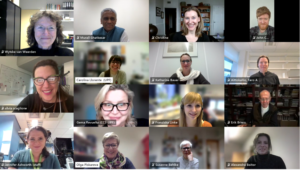
The kick-off meeting of Mac4Me partners, Feb 2025
We are very proud to train two out of 18 Doctoral Candidates, building upon the expertise of Drs Ian Woods, Adrian Dervan and Prof Fergal O’Brien in biomaterials and 3D bioprinting and Dr Olga Piskareva in neuroblastoma biology and 3D in vitro cancer models.
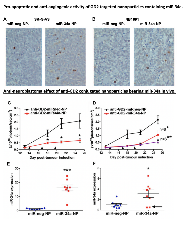
I’m excited to kick off my second-year PhD journey with a deeper dive into cancer research. This is my first blog post of the year, and I’m eager to share what’s sparking my curiosity. So, I came across a paper by Tivnan et al. (2012), which focused on the targeted delivery of microRNA-34a (miR-34a) using nanoparticles. What intrigued me most was how these nanoparticles are designed to deliver therapies straight to cancer cells. Neuroblastoma is a highly aggressive and difficult-to-treat tumour, so finding a way to target it without affecting healthy cells could be a breakthrough.
Here’s what makes this study so exciting: the team developed a nanoparticle system coated with anti-GD2, a molecule that recognizes and binds to GD2, a marker commonly found on neuroblastoma cells. Think of these GD2-coated nanoparticles as specialized delivery trucks with a precise address—they’re designed to deliver miR-34a.
Now, let’s dive into the details of miR-34a’s role. MiR-34a isn’t just any therapeutic agent—it’s a master regulator capable of influencing multiple genes involved in cell growth, survival, and blood vessel formation. By releasing miR-34a into tumour cells, this study activated pathways that induced cell death and suppressed angiogenesis, preventing the tumour from forming new blood vessels. It’s almost as if miR-34a is a conductor orchestrating a complex, multi-step attack on cancer, using the tumour’s own cellular mechanisms against it.
The Results? A Direct and Multi-Layered Attack on Tumor’s
In their mouse model, the GD2-targeted nanoparticles packed with miR-34a significantly reduced tumour growth. These “smart” nanoparticles didn’t just shrink tumors by inducing apoptosis (cell death); they also cut off the tumor’s blood supply by promoting the expression of TIMP2, an anti-angiogenic protein. Essentially, the tumor cells were directly targeted and deprived of the resources they needed to survive—a powerful one-two punch.
Where Do We Go From Here?
This study is an excellent example of how targeted therapies could evolve to tackle other types of cancer. Traditional therapies, like chemotherapy, often affect both healthy and cancerous cells, leading to significant side effects. In contrast, this targeted approach delivers miR-34a specifically to neuroblastoma cells, which could be especially beneficial for pediatric patients who need treatments that minimize harm to developing bodies. Imagine pairing nanoparticles like these with different therapeutic targets, such as GPC2, ALK, or PDL1, or even combining them with existing treatments to boost effectiveness while minimizing side effects. For those in the field, the potential here feels like a breakthrough waiting to happen.