How do researchers study cells? How do we get the nitty gritty?
We use many methods to tag and chase various cell components. One of my favourites is fluorescent microscopy. It allows the use of nearly all spectrum of colours from blue to purple in one go. However, we prefer to narrow it down to 2-3 colours and avoid their overlap.
How does it work? First, we use DAPI or Hoescht, which are blue fluorescent dyes used to stain DNA. This way, we tag the nucleus of the cell. Then, we tag a protein of interest. In our case, it was MYCN, a protein that acts as a transcription factor. MYCN amplification is associated with poor prognosis in neuroblastoma. As a transcription factor, it binds to genomic DNA and is located in the nucleus. We used a specific antibody that was labelled with a green fluorescent dye. Look at the image below. The green colour pattern overlaps with the blue colour. Then, we tagged the cytoskeleton, a complex of various proteins that hold the cell architecture and dynamics. We used phalloidin with red fluorescence. It is a highly selective bicyclic peptide and a popular choice for staining actin filaments.

Now, we can enjoy visualising cells and test different research questions. For example, how do cells respond to a drug? Or how do neuroblastoma cells spread?
Written by Olga Piskareva






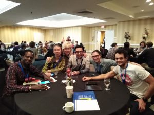
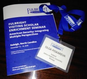 The theme of the seminar was American Security: Integrating Multiple Perspectives. With the great support from North Carolina University, we stepped out our comfort zones and explored security topics through lenses of our cultural backgrounds and life experiences. Food Security, Energy Security and Environmental Security. Eighty-four Fulbrighters from almost 40 countries were sharing their stories on how these issues are dealt with in their home countries! We learnt to talk through being open-minded, find things in common and come to a balanced solution. This is not a-one-size-fits-all solution. We have more in common than we have thought. We have become friends and partners who build bridges and connect people and countries. The Fulbright Programme helped us to realise it!

 On November 22, almost all Americans and visitors celebrated

 Barnes & Noble bookstore is located in the former Power Plant. The features are easily spotted. From outside, the building looks like a Plant for modern social activities. Ugly slightly, isn’t it? Though, it is a different feeling when you enter the bookstore. The Plant scaffolds, chimneys and pipes are nicely crafted into a warm welcoming environment. Even lights are dimmed as back then. Rambling through the bookshelves and feeling the magic of the place and unread stories on them. You can pick up a book, sit where you are and enjoy the reading. Maybe it is the feeling of my childhood full of books and hours of reading?

