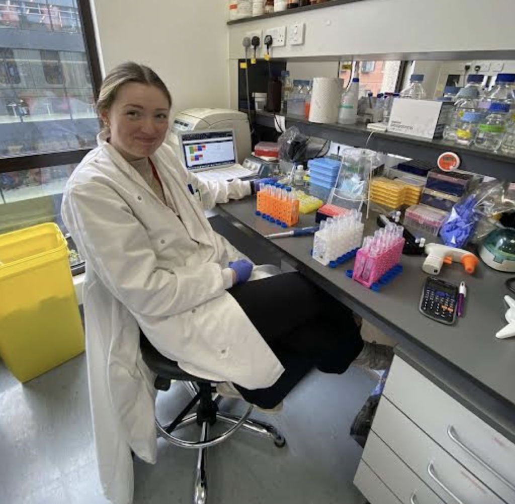
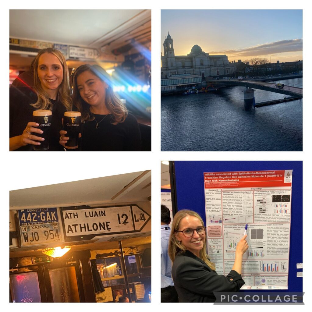
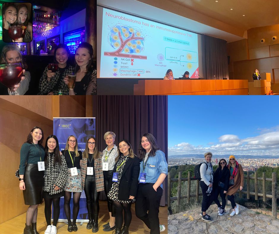
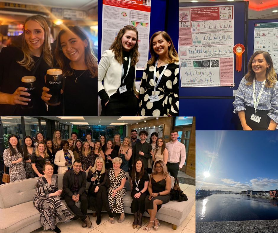
In-person conferences are back at last! In March 2021 I attended the IACR conference for the first time, albeit virtually. While there were some great talks at IACR 2021, the virtual experience was lacking in the networking and socialising opportunities that go hand-in-hand with traditional conferences. So I was very excited to be Cork-bound for IACR 2022 in March of this year.
To my surprise, my abstract was selected for a Proffered talk, meaning I had 10 minutes in the limelight of the IACR podium to present my research on immune markers in neuroblastoma. Having gone two years without presenting to a crowd, it was an adrenaline-filled experience, and it was great being surrounded by my colleagues after the talk rather than being at home alone in front of my computer.
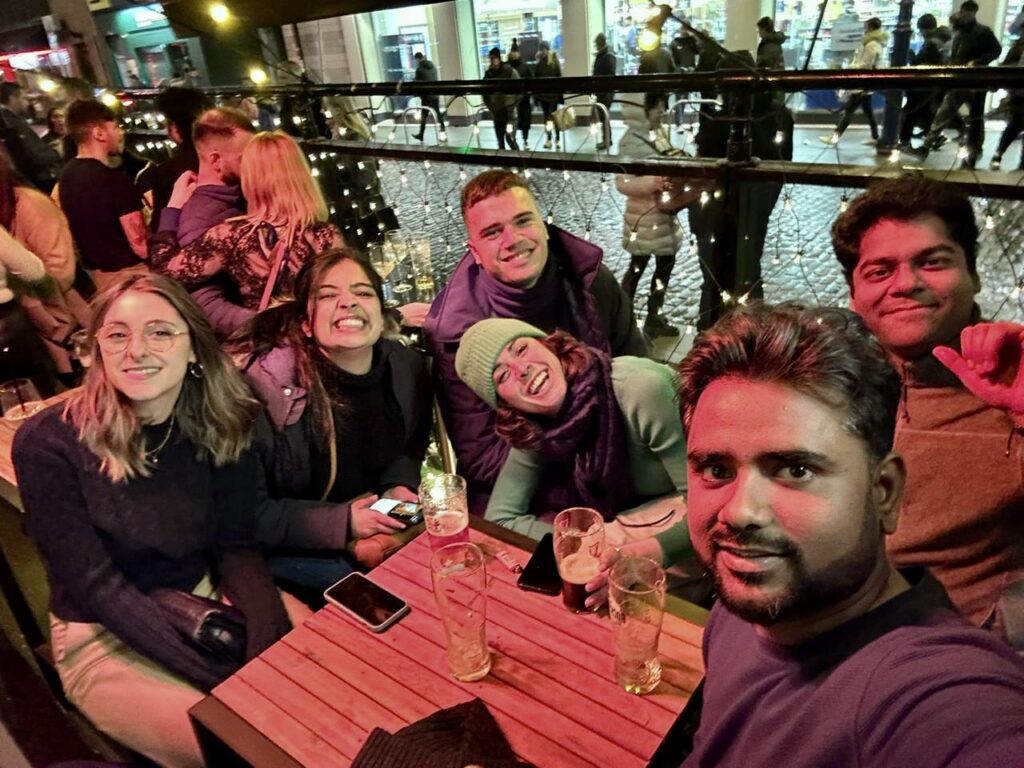
There were many very memorable research talks and posters at IACR, but some of the best memories came from the moments in between the scientific sessions. From the train down to Cork with my lab group, to buffet dinners, a quick journey into Cork city, going for a swim in the lovely hotel pool, and singing and dancing the night away at the gala dinner on the last day of the conference.
One of the highlights of the conference for me was the awards ceremony at the gala dinner, where to my delight I was awarded the Best Proffered PhD talk! What a fantastic way to end a great few days at IACR 2022.
It was February 2020, just before one of the biggest global pandemics struck, that I attended the IACR as a research assistant. It was my first official conference and it is safe to say ‘Imposter Syndrome’ was my main feeling going down to Galway on the train. Fast forward 2 years and my feelings travelling to IACR 2022 in Cork could not be more different. It is amazing what starting a PhD during a pandemic can do for your confidence and skills as a researcher – a sink or swim moment if there was ever one. My first IACR in Galway was one to remember surrounded by like-minded scientists, all brimming with new ideas and exciting discoveries. As such, I had high hopes for IACR 2022. And it did not disappoint.
My PhD project focuses on the development of a vaccine to treat neuroblastoma so I was very excited to hear talks from some of the leading experts in vaccine research, both in industry and academia. I gained so much from hearing these experts discuss their research but also discussing other important topics like career progression and how to keep a work/life balance in research. It was refreshing to hear that as scientists we don’t have to (and shouldn’t) work ourselves to the bone 24/7 to be successful. As a young scientist planning to continue into academic research, this left a lasting impression on me. To top off what was already a hugely beneficial conference for me, my poster was shortlisted for a prize. I was shocked, delighted and excited all-in-one. Starting my PhD during a pandemic was not without challenges. Delays in deliveries, delays getting trained on equipment and multiple lockdowns led to what felt like (for me) quite a disjointed start. For my research to be shortlisted by experts was, to be honest, a relief. To know that my work stood out was extremely important to me and that all the hard work does pay off. When my name was called out at the Gala dinner as a Poster Prize Winner, all the doubts that I had (doubts that we all have as scientists) disappeared. I felt very proud and very grateful that my research was recognised at that level. There is no doubt that in-person conferences give a huge boost to young researchers, and I really look forward to presenting my work at the next IACR meeting.
At the beginning of my career, I worked for two years in a Ukrainian company organizing international industrial conferences. So I have insider knowledge of how the conference works, and that the determining factor for the success is the active communication between the participants. And at the RCSI research day and Cork IACR conference, this component was perfect. At both events, I presented my poster and had a chance to discuss the recent advances in neuroblastoma epigenetic drug research. During RCSI Research day, I was excited to learn about the accomplishments of other undergraduate studies and was thrilled to learn that my classmate is participating in research too. He had developed an online recourse to practice cardiac auscultation, which is extremely useful for my medical studies. But professionally, I enjoyed the cancer research posters and presentations at the IACR conference and was eager to meet the researchers working on medulloblastoma, a paediatric neural cell cancer, and the research team from UCD, the neighbours of our university who worked on breast cancer. It was the most valuable opportunity to take a glimpse into other research, become inspired by the most ingenious methods, and cultivate professional knowledge and personal connections – I am so lucky I have been at RCSI Research day and the IACR conference! I have greatly enjoyed my time, and I am looking forward to (hopefully) going to the next year’s conferences again.
It is time for a full group presentation here at the blog! Throughout the month we shared about our group members and their research focus on Twitter. Now, we would like to share more about the group here and invite you to keep following us on social media.
The Cancer BioEngineering Group is a research group led by Dr Olga Piskareva at the Royal College of Surgeons in Ireland. The group has 6 PhD students developing research projects around neuroblastoma biology.
Our projects address topics related to neuroblastoma microenvironment, cell interactions, tumour resistance and the development of new therapies. To do that we use 3D in vitro models, identify immunotherapeutic targets and evaluate extracellular vesicles.
We are a dynamic group proud to be engaged in research, science communication and patient involvement. We do that through different initiatives.
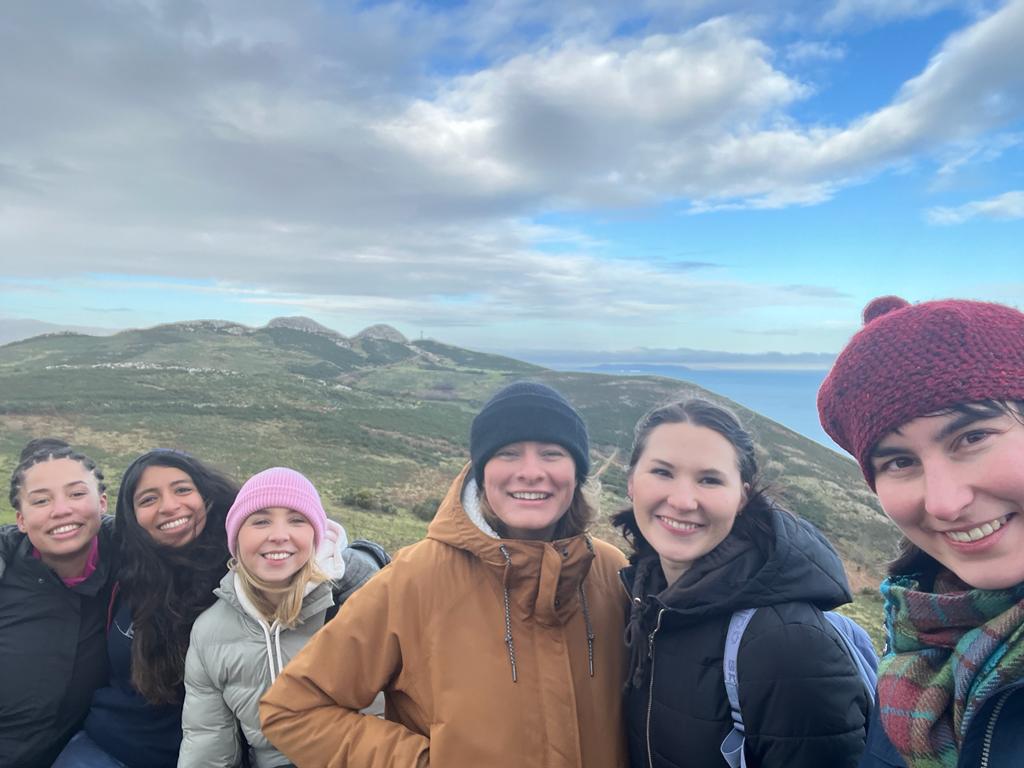
We promote neuroblastoma awareness through different activities. For instance, last September at the Childhood Cancer Awareness month we promoted a hiking challenge to raise money and increase awareness of neuroblastoma. We hiked for 30km at Wicklow mountains in a day and raised over € 2,000 for neuroblastoma research charities.
We are also present in social media, creating content in the form of blog posts and tweets to share the science we are doing.
We are always happy to answer questions and interact with the public. Follow us on our social media channels and read our blog to know more about us and our research.
Thanks for reading and we go ahead with neuroblastoma research!
Understanding how tumour cells interact with the other cells in the body is crucial for an effective treatment. Moreover, it can help to identify patterns that are exclusive of tumour cells to be a target in treatment.
The interactions of tumour cells with the surrounding tissue, the microenvironment, affects chemotherapy sensitivity, immune cells recognition and expression of molecules on the cell surface, to only cite a few interferences.
This is particularly crucial in metastatic cells, which are cells that have spread to other parts of the body coming from the primary tumour location. Specifically, for neuroblastoma half of patients with high-risk disease present a metastatic tumour at the diagnosis. In addition, one of the organs that are mostly populated by metastatic neuroblastoma cells is the bone marrow.
A review paper recently published address some important aspects about the interactions between neuroblastoma cells, bone and bone marrow resident cells1. This review argues in favour of understanding these interactions to search for new targets for therapy.
However, neuroblastoma cells proved to be difficult to characterise due to dynamic changes induced by external stimuli. Therefore, neuroblastoma cells change upon exposure to the bone marrow microenvironment.
The authors present some studies showing that neuroblastoma cells infiltrating the bone marrow express receptors for small proteins called chemokines that induce cell adhesion in the bone marrow. On the contrary, the cells did not present on their surface molecules that stimulate the immune system recognition. Therefore, they are naturally invisible to the action of this system.
Moreover, it has been shown that metastatic tumour cells release extracellular vesicles expressing GD2. These vesicles have an important role in cell-cell communication and the GD2 is a marker exclusive of neuroblastoma cells. Thus, it facilitates the identification of metastatic cells.
These alterations on neuroblastoma cells surface after they interact with bone marrow cells may facilitate the invasion and spread of the tumour. Thus, looking closely to that may help to develop more effective treatments for neuroblastoma.
At the Cancer Bioengineering Research Group, many of our projects are related to tumour resistance, cell interaction and the tumour microenvironment. These three aspects are very important to understand neuroblastoma at the tissue level. We study them and expand this research to applied projects aiming at the development of new therapeutic modalities.
For instance, we are currently evaluating the effect of extracellular vesicles from different neuroblastoma cell lines in the induction of proliferation and increased viability. Moreover, we are studying the interaction of neuroblastoma cells with immune cells such as macrophages. Finally, we are also identifying targets to develop an anti-tumour nucleic acid-based vaccine against neuroblastoma.
We go from basic to applied research interconnecting the findings and expanding the understanding of neuroblastoma biology. Ultimately, we aim to improve treatment and quality of life for patients.
Written by Luiza Erthal
References
1. Brignole, C. et al. Bone Marrow Environment in Metastatic Neuroblastoma. Cancers13, 2467 (2021).
Looking carefully we can easily see that children are very different from adults. They have different needs, desires, likes and dislikes. Not surprisingly, the children body is also very different in their functioning and response to medical needs. Therefore, cancer in children has many different characteristics when compared to cancer in adults. Childhood cancer is different in terms of the most common types, the causes, the treatment and the course of the disease.
Firstly, childhood cancer is rare and this sometimes impairs an early diagnosis. Therefore more aggressive diseases tend to be present at the time of diagnosis. Nevertheless, there are specific types of cancer that are more common in children, which helps in the diagnosis. They are cancers affecting the blood and lymph nodes (leukaemia and lymphoma), the brain (astrocytoma), the liver and the bones (osteosarcoma). These types of cancer are less common in adults.
Another important difference between adult and childhood cancer is the leading cause of the disease. Most of the time the cause of childhood cancer is unknown, although genetic contributions related to overexpression or deletion of genes can be determined. On the other hand, adult cancers are frequently associated with alterations in the DNA (mutations) as well as lifestyle.
The treatment plays an important role in the differences between adult and childhood cancers. Usually, similar treatments are used for both adults and children, including chemotherapy, radiotherapy, surgery, transplants and immune therapy, according to the type of cancer and its stage. However, the doses and types of drugs may differ between them. The differences in the treatment go beyond the doses and encompass the mechanisms of action and possible long term toxicities of drugs. For example, the use of drugs that damage DNA can be prohibitive in children due to the increased risk of secondary cancers in the future.
In conclusion, specific types of cancer are more common in children and the cause of this disease is frequently unknown. Fortunately, children have great possibilities to survive cancers but the treatment needs to be carefully chosen and its long-term effect on the body have to be monitored for their whole life.
Written by Luiza Erthal
References
Kattner, P. et al. Compare and contrast: pediatric cancer versus adult malignancies. CancerMetastasis Rev. 38, 673–682 (2019).
The determination of the tumour stage is an important step after a neuroblastoma diagnosis. The stage of neuroblastoma is determined depending on tumour location and if it has spread to other parts of the body. This will guide risk group assignment and treatment choice.
The first staging system for neuroblastoma, the International Neuroblastoma Staging System (INSS), was developed in 1986 and is based on the pathological evaluation of the tumour after a removal surgery. In 2005, The International Neuroblastoma Risk Group Staging System (INRGSS) started to be used. This system is based on tumour images before any surgery. Therefore, it is based on image-defined risk factors to determine the tumour stage (see table below). It also uses clinical, pathologic, and genetic markers to determine the risk groups, which can be low-risk, intermediate-risk, or high-risk.
Reference: Neuroblastoma – Childhood: Stages and Groups, Cancer.net.
Recently, the Children’s Oncology Group (COG), a clinical trial group dedicated to paediatric cancer research revised the classification system they use to determine tumour stage for enrolment in clinical trials1. Previously, they have been defining the tumour stage based on the INNS system. Now they proposed a revised classification that takes into account the INRGSS and chromosomal alterations.
Key clinical and biological factors used in the neuroblastoma risk classification include age at diagnosis, disease stage, tumour tissue appearance under a microscope (histology), the status of the gene MYCN that affects tumour growth, the amount of DNA in a tumour cell (called tumour cell ploidy), and alterations in the DNA.
They analyse the outcome of almost 5,000 patients to define risk groups based on the INRGSS, using alterations in the DNA of tumour cells as a biomarker and considering current therapy modalities. In general, they found that the correlation of stages between systems is not exact. However, the differences in survival were minimal when comparing staging systems, which corroborates the use of the revised version.
In general, the new version classifies L1 and L2 tumours as low risk, except for L1 tumours with alteration in the gene MYCN and that cannot be removed by surgery, which is high-risk. For L2 tumours, MYCN status and age can be used to evaluate prognosis. Stage M tumours can be classified as high risk or intermediate-risk depend on age, MYCN status and DNA alterations. In conclusion, low-risk groups have excellent outcomes with any or limited therapy, the intermediate-risk group have very good outcomes and high-risk groups have inferior outcomes despite therapy.
This new version of the COG classifier will provide a uniformization of patient risk classification for clinical trials, ultimately enabling the comparison between different trials.
Written by Luiza Erthal
Reference:
1. Irwin, M. S. et al. Revised Neuroblastoma Risk Classification System: A Report From the Children’s Oncology Group. J. Clin. Oncol. JCO.21.00278 (2021)
For most neuroblastoma cases a tissue biopsy, which means remove a piece of the tumour and analyse under a microscope, is performed to confirm the diagnosis, allow the risk stratification (low, intermediate or high-risk cancer) and determine treatment. However, it is not always possible to perform a biopsy. In these cases, other tests are available such as ultrasound, x-ray and urine test1. Although these tests help the diagnostic, they are much less informative to determine the risk group and to guide treatment.
Cancer is a genetic disease and as such several genetic alterations are present in the DNA and can be useful to determine a diagnostic and prognostic of the disease. These alterations can also be useful to monitor treatment effects. But, how we can examine the DNA without using a piece of the tumour?
The answer for that relies on our blood. Cancer patients have cancer cells circulating in their blood and these cells release what we call cell-free tumour DNA. The level of cell-free tumour DNA in the blood of a healthy individual is low while this is increased in cancer patients. The detection of cell-free tumour DNA with specific alterations may help to not only detect the disease but also determine if the tumour cells are changing after treatment.
Recently, a trial for a blood test that can detect 50 different types of cancer was launched in the UK2. The test called the Galleri test look for cell-free tumour DNA in the blood using modern genetic sequencing technology. It spots the DNA that has changes common in specific cancers but not seen in healthy cells.
One of the main questions of this trial is if the test can find early stages of cancers. Although neuroblastoma is cancer that could benefit from this test, unfortunately, it is not one of the cancer types detected. However, the presence of cell-free tumour DNA was already detected in neuroblastoma patients’ blood3. Moreover, several alterations in the DNA of neuroblastoma patients have already been reported to have predictive value for disease progression and treatment monitoring. These alterations include the increase in the number of specific genes, genes breakdown or increased activity of certain genes. Their presence may indicate poor outcomes and early detection could guide to specific treatment options.
The big challenge is to have a non-invasive diagnostic method, such as blood tests, that are sensitive enough to detect the early stages of the disease. Specifically for neuroblastoma, comprehensive analysis in clinical trials regarding cell-free DNA levels and their specific changes over time would help to advance the development of liquid biopsies, such as blood tests, for this type of tumour.
We are launching a new initiative #AskLuiza to help the public and patients know more about advances and current trends in neuroblastoma.
Luiza is a research writer at the Cancer Bioengineering Research Group. She holds a PhD in Biomedical Sciences from Trinity College Dublin. You will ask a question and Luiza will look for the answer in peer-reviewed research papers that the research community trust.
And the story began with a meeting of fantastic 7 at the very beginning of Dublin Mountains Way in Tallaght at 6.30 am on September 25th. The spirit, cheer, backpacks with essentials and branded tops were on, Strava was launched and we swiftly headed off.
It was quiet, dark and cheering. No one was on the streets, a few cars passed by. We took towards Bohernabreena reservoir through the sleepy estates of Tallaght, sensing the sunset. Clouds were low and the highest peaks in the Dublin Mountains including Seefingan, Corrig and the highest, Kippure were in the mist. Nevertheless, we were full of energy and hopes to see it later.
Cheat chats and jokes were here and there, we walked in small dynamic groups recalling our pre-covid life and stories that happened during the lockdown. A mix of newbies and maturating research students. We met some in person for the first time since the COVID restrictions admitting that our visual senses are extremely important to memorise a person and recognise him/her on the next occasion. We were enjoying this face-to-face communication and our team re-connection.
The first 8 km flew in a flash. We stopped for our breakfast in Dublin Mountains. The grass was wet, the sky was blue. Mountains started to draw their shape through the clouds. Yoghurts, fruits, bars immediately disappeared in our stomachs. Everyone was happy to lighten their backpack. Every little helps!
A few plasters were glued, and we continued on at a very good pace. The sky was changing with sunny spells. We travelled around Spinkeen and Killakee at their base doing up and downhills and verifying our route with the hiking app. At the 20 km mark, we stopped for lunch. Sandwiches, grapes, mandarines and sweets were shared and eaten and then polished with chocolates from the recent Nadiya’s home trip. Jellies left untouched.
At 25 km, our blisters reminded us of being humans. Our pace slowed down and we started a very mild ascent to Tibradden Mountain leaving the Pine Forest or Tibradden Wood behind. We climbed further to Fairy Castle, the highest point on the Dublin Mountains Way (537m). Throughout the entire way, Dublin showed its best views of the Phoenix Park and the Pope Cross, house roofs, Aviva Stadium, two Chimneys, Dublin Port… The scenery was fascinating and breathtaking. We saw Howth and Dun Laoghaire, Sugar Loaf… We met groups of Germans, French, Irish and many others.
At Three Rocks Mountain/Fairy Castle, we started our descent and entered Tiknock forest. This part was steep. We crossed the Gap Mountain Bike Adventure Park to reach Glencullen. Got lost at the end but just for a sec and reached the Glencullen junction at 2.30pm. It took us 8 hours with walks and stops from start to finish to complete the 30 km challenge in a day. We got tired but felt happy and satisfied.
We aimed to raise awareness of childhood cancer in general and neuroblastoma in particular as well as honour children with cancer, their parents, siblings, friends and careers, doctors and nurses, volunteers in the hospitals and researchers working to find cancer weaknesses and develop new treatments that are friendly to patients and target cancer aggressiveness.
We will count our tally in the coming days and transfer it to three wonderful charities that support childhood cancer research.