One Day of the Life as a Researcher: summer undergraduate student

This summer I worked under Dr Piskareva supervision in the remote research program. My original plans for a lab experience were put down by COVID. My ultimate goal was to write the review article on the potential uses of retinoic acid in neuroblastoma research.

Nadiya Bayeva

Before starting the project, I didn’t have any specific expectations. While I always had fun picking the primary articles apart and summarising the gathered data during my undergrad and med school, I didn’t believe that this experience would be the special one. And I kept thinking so as I was collecting the information from the numerous data on cell cultures. And as I was looking at the mice models studies. And clinical trials.

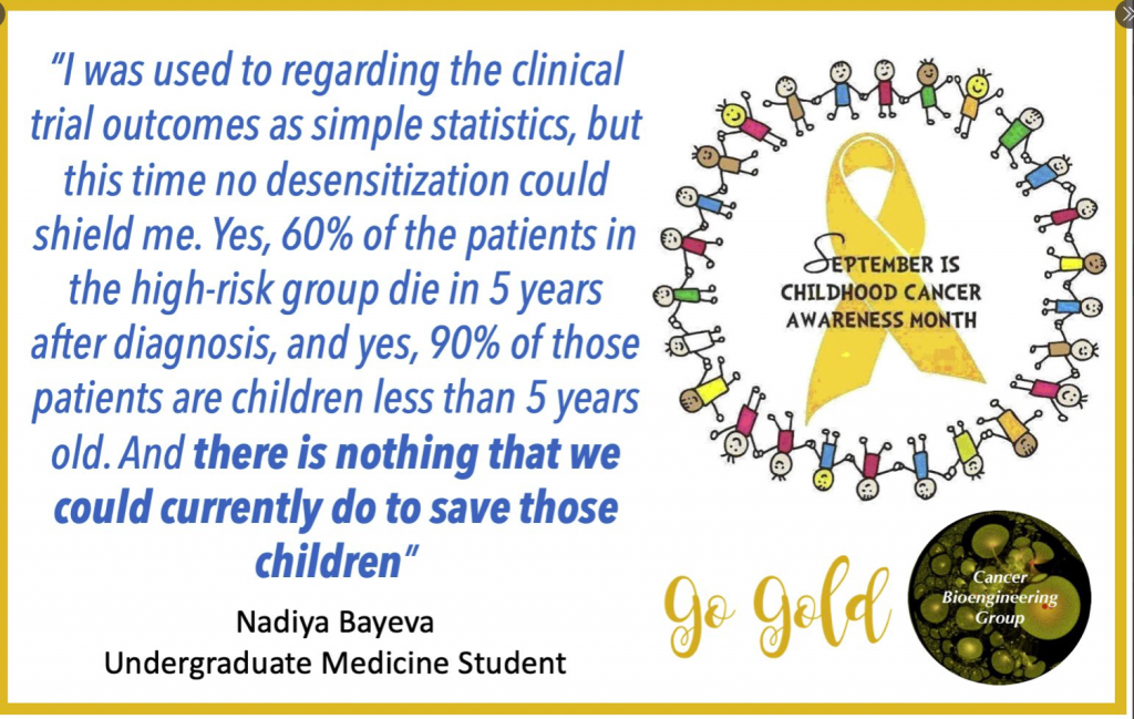
Then I started to write my introduction, and so researched the information on the neuroblastoma prognosis, contemporary treatment protocol and outcomes. And suddenly I saw my project in the new light. I was used to regarding the clinical trials outcomes as simple statistics, but this time no desensitization could shield me. Yes, 60% of the patients in the high-risk group die in 5 years after diagnosis, and yes, 90% of those patients are children less than 5 years old. And there is nothing that we could currently do to save those children.

On the other hand, this realisation brought meaning to my work. This time, I was not doing a PubMed search to get a good mark or CV reference. Instead, I was looking for the possible treatment of the disease. My review on the current knowledge about retinoic acid will let the other primary researchers target the most promising drug for future experiments and, eventually, create a novel and effective therapy to help those children.

And isn’t it what we are all striving for in medical research?

International Childhood Cancer Awareness Day – February 15th 2019

International Childhood Cancer Day (ICCD) was founded in 2002 by Childhood Cancer International (CCI). Each year on February 15th we unite together to recognise childhood cancer as a national and global child health priority & to raise support, funding and awareness of this devastating desiease.

This year we team up with Amorino to run Hot Chocolate Morning.  Please come along! All proceeds go to CMRF/NCRC and CFNCRF.

If you can’t join us, you can simply follow the link and donate ‘a cup of coffee/hot chocolate’ to CMRF Crumlin, the Conor Foley Neuroblastoma Research Foundation & Childhood Cancer Foundation


Scientific part of my journey

Reading my posts, it looks like I am more enjoying the cultural part and almost forgot the main reason I crossed the Atlantic with the Fulbright wings.

The first month in the lab was more a warming up. Where is my desk? Where is the cell culture rooms? How do they run it? How different is it? So, many microscopes – am I capable of imaging? And so on and so forth…

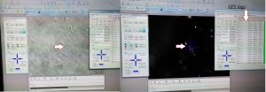
My typical day starts at 8-8.30 am and finishes once all is done. It may be 6pm or 10pm. Once the experiment is set up, I have to monitor cells every 24 hours for 5-7 days with no weekends or days off. The monitoring includes imaging. Lots of imaging. Every condition has 20-30 single cells to follow up. Each cell gets its own GPS tag manually to be able to image exactly the same cell as it grows and becomes a group of hundreds by multiplication. For example, I am running 8 different cell lines in 3 experimental conditions. So, 20-30 cells per all 24 combinations give us 480-720 individual cells to follow up. The imaging takes ~5 hours every day. After 5 days, I will have 2400 – 3600 pics of cells to analyse. It will be fun! I may need lots of Guinness to fly through that numbers.

Tagging cells. The left arrow points to a group of neuroblastoma cells. The arrow in the middle point to the same cells, but this image allows you to see the actual number of the cells. This group has 8 cells. The right arrow points to individual GPS tags for each cell

At the next step, I will select some of the conditions for video recording to trace cell fate from a single neuroblastoma cell to a metastatic niche consisting of hundreds of them. This video will show me how it all happens minute after minute.

Is not it exciting? I am thrilled!


Research Summer School 2018

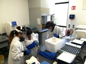
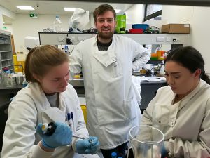
Another year, another Research Summer School students. We are hosting 4 students (Jessica, Dawn, Dola, and Jeff) this year. Some of them will be medical doctors, another will do research after the graduation. For them, the 8-weeks lab placement is a window into the reality of the everyday science. How cancer cells look? How do they grow? Where do we store them? How do we know that we have identified a new drug or a new target to study further? Do researchers have a sense of humour? Do they like donuts?

Why do they wear these astronaut helmets?

We have already said Good Bye to Jessica. Dola and Dawn’s projects are coming to an end this week, while Jeff is staying till the end of August.

How current IT advances help in research?

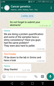
Here is the perfect example of the teamwork troubleshooting protein extractions. My Dream Team 2018 in action. The current information and communication technologies allow to stay connected and respond quickly.

Five minutes later in the lab: troubleshooting is the exchange of experiences!


A new 3D strategy to study neuroblastoma

Our body has 3 dimensions: height, width and depth. Every single part of our body grows in the same 3 dimensions. This is true for cancer cells. Researchers use different ways to study cancer cells behaviour, how they grow and spread. We grow cells in the flasks, where they change their structure and shape and become flat losing one dimension. This is a very popular approach. We also grow cells in mice, where cells keep their 3D shape and mimic their behaviour to one observed in humans.

It is well known that we need to give a different amount of drug to kill cancer cells grown in flasks and in mice. This, in turn, delays the development of new drugs. Why does it happen this way? So, the drug works only on one side of the cell when they grow on the flat surface. In contrast, in mice, drug surrounds the cancer cell habitat and attacks cells at the edge first and then getting to those at the core. So we need more drug to kill cancer cells in mice.

We decided to design a new way to grow cancer cells that recreate their growth in 3 dimensions as in the human or mice body. We used special cotton wool like sponges as a new home for cancer cells and populated them with cancer cells. At the next step, we gave cells the drug at the different amount and checked what happened.

To understand cell fitness we stained them with red and blue dyes. On the left bottom side of the image, we see an equal amount of red and blue dyes telling us that cells were healthy and fit. Cells did not get any drug. When we gave a little amount of the drug but enough to kill cells in the flask, the balance of red and blue dyes was the same telling us that nothing really happened (the image in the middle). Cells were feeling well and healthy. The right bottom image has only blue dye. In this case, cells were given the amount of drug enough to destroy cancer cells in mice or humans. The lack of red dye tells us that this time the drug worked and killed the cancer cells.
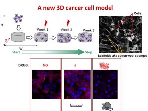
We found that the drug killed cells on sponges only at doses enough to do the same in mice.

So, we concluded the new tactic to grow cancer cells in 3D on cotton-like sponges can bridge the gap between traditional way and animal models. This new strategy to grow cells on sponges should help to understand cancer cell behaviour better and accelerate the discovery and development of new effective drugs for neuroblastoma and other cancers. This, in turn, will make the outlook for little patients better and improve their quality of life.

This work has been published in Acta Biomaterialia and presented recently at the Oral Posters Session at the 54th Irish Association for Cancer Research Conference 2018.

This study was supported by Neuroblastoma UK and National Children’s Research Centre.

You can find more at

A physiologically relevant 3D collagen-based scaffold–neuroblastoma cell system exhibits chemosensitivity similar to orthotopic xenograft models.

IACR Meeting 2018 Programme
How is it feeling?

The fact of being shortlisted is very encouraging. It means that my research proposal and the career achievements fit the merit of this award.  No doubt it was fantastic experience overall, not often the shortlisted candidates have an opportunity to speak for themselves.

How is it feeling after the interview?… Well, I do not have a firm answer… It is a big difference when you explain yourself in written and spoken forms… No chance to edit your real-time talk… How many times have that 30 minutes played back and re-run in my head? I lost the count… Did I bring the point across? Did I do things in right time and at a right pace? Should I have structured the answer differently? Each re-run brings new ways to answer the same questions, indeed, in a better and more concise way…   Having the mind that is constantly analysing the situation is not helpful.

Think, the competition was very tough, and only 1 in 10 made to the 2 days interview for the Fulbright Award (maybe the ratio even higher). Twelve candidates were interviewed yesterday and the same numbers are to be today.  What are the chances to get to the final? I have to wait until March… and meanwhile, keep applying for grants and doing something meaningful.

My next stop is at the Irish Cancer Society this Thursday to film a short video about my research and neuroblastoma challenges. The video should be available for the International Childhood Cancer Research Day on February 15th.

The First Big Challenge in New Year

The first month of the new year and the first challenge. Monday is the big day for me. In the essence, my grant application was shortlisted for the interview where I have to face the challenge and prove that I worth it.

Anyone for a challenge?

The proposed application seeks to go to the US lab and gain an expertise in an interdisciplinary methodology to monitor and capture the dynamic of cancer spread (metastasis) in real time. This experimental approach would accelerate our understanding of neuroblastoma metastasis which is one of the reasons for failure in the treatment of neuroblastoma. If we know how neuroblastoma cells spread, then we can find the weaknesses in the process and create new drugs or use existing to target it.

I feel that sharing my worries with you makes me stronger. I am looking forward to this challenge with my head up and hope to feel your support at this crucial moment.


Networks of Galaxies

Some researchers look into possibilities to predict real “me” by analysing what and how was said on social media.

This network of galaxies is a new project to get insights on how and where childhood cancer is placed on social media. It is a collaborative project with Prof Richard Arnett. It is already very exciting and more to come!


What is the risk group classification system?

To be able to guide the treatment of neuroblastoma patients, doctors have developed a number of classification systems. Although sharing common features, they slightly vary by medical center, country and continents making direct comparisons of treatment results difficult. Doctors and scientists are trying to consolidate all systems in one in order to evaluate treatments in the past, currently ongoing and in the future.

Scientists have suggested a newer risk group classification system, the International Neuroblastoma Risk Group (INRG) classification that would incorporate the best knowledge gained and recent advancements in the disease imaging and neuroblastoma molecular diagnostics. This system is based on imaging criteria using the image-defined risk factors (IDRFs) and the prognostic factors such as:

  • The child’s age
  • Tumour histology (the tumour appearance under the microscope)
  • The presence or absence of MYCN gene amplification
  • Certain changes in chromosome 11 (known as an 11q aberration)
  • DNA ploidy (the total number of chromosomes in the tumour cells)

The table is adapted from Pinto NR J Clin Oncol. 2015

Using these factors the INRG classification put children into 16 different pre-treatment groups (lettered A through R). Each of these pretreatment groups is within 1 of 4 overall risk groups:

  1. Very low risk (A, B, C)
  2. Low risk (D, E, F)
  3. Intermediate risk (G, H, I, J)
  4. High risk (K, N, O, P, Q, R)

This system has not yet become common across all medical centers, but it is being researched in new treatment protocols.

Doctors and scientists are planning to improve the INRG classification system by incorporating other molecular diagnostics data such as profiles of the neuroblastoma genome (DNA), transcriptome (RNA), and epigenome* in order to make precise prognostication even better.

* The epigenome is made up of chemical compounds and proteins that can attach to DNA and direct such actions as turning genes on or off, controlling the production of proteins in particular cells

References:

Pinto NR, Applebaum MA, Volchenboum SL, Matthay KK, London WB, Ambros PF, Nakagawara A, Berthold F, Schleiermacher G, Park JR, Valteau-Couanet D, Pearson AD, Cohn SL. Advances in Risk Classification and Treatment Strategies for Neuroblastoma.J Clin Oncol. 2015 Sep 20;33(27):3008-17.