Hot off the Press!

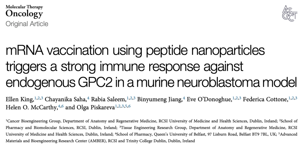
We are over the moon with our work being recognised by the American Society of Gene & Cell Therapy Molecular Therapy Family of Journals. The results are published in a Special Issue focused on the Advances in Paediatric Cancer Therapy. It is a true milestone in a challenge we undertook in collaboration with Prof Helen McCarthy at Queen’s University Belfast, with High-Risk, High-Gain funding from the Higher Education Authority North-South Research Programme. We did prove the HEA Ambition to highlight the huge potential of their initiative. Now, I can share the score for our application – it was 99/100*! Oh, my!!!! We are so happy that our idea was recognised back then. *I am still struggling to reach this threshold in other applications. 😉


Indeed, great research is teamwork, trust and collaboration – huge appreciation goes to Chayanika Saha, Federica Cottone, Eve O’Donoghue, Rabia Saleem, and Binyumeng Jiang. You are the Rising Stars!

Ellen receives her IACR/EACR Junior Scientist Award 2025


However, my extreme credit goes to Ellen King, PhD, who took on my ambitious challenge (go/no-go!), trusted me, went above and beyond, and turned her PhD project into this publication with passion and diligence. Just for context, it took us 12 months with 7 rejections to receive the quality approval mark for this publication. All this time, she was on top of the reviewing and publishing game after graduating with her PhD in 2024, developing her own immuno-oncology portfolio in London. Onwards and upwards Ellen King, PhD!


We are proud to collaborate with the family of John Foley CMC, FIMCA, MIET. – the true inspiration for not giving up! Their story of the neuroblastoma battle is heartbreaking and inspiring at the same time.
The Higher Education Authority funding helped us to secure follow-up funding from the Health Research Board (HRB) & the Conor Foley Neuroblastoma Cancer Research Foundation ( via HRCI – Health Research Charities Ireland) and Neuroblastoma UK. We appreciate their trust in our ambition and vision. THANK YOU!

The VHI Mini Marathon 2026 – the Start of our Childhood Cancer Awareness Campaign. 

Yesterday, 30,000 runners, joggers, and walkers took part in the 44th VHI Women’s Mini Marathon. We were ages 14 to 92, from diverse cultural, educational, and fitness backgrounds. Each set their own ambition and target. It was my fourth race. My targets stayed the same: raising awareness for Childhood Cancer Research, supporting the Conor Foley Neuroblastoma Cancer Research Foundation, and finishing within 1 hour and 15 minutes with a smile.

The atmosphere was cheering and empowering, and the weather was very kind to us. I was in a pink wave, and we started about 12:30ish. Running in a big company with your allies is enjoyable and empowering. I was sinking into the diversity and variety of running women and supporters along the road, enjoying every minute. Some took over me, and I took over some. Somewhere after 5K, I was tapped by my TERG colleagues, so the race became even more enjoyable.

The Vhi Mini Marathon 2026 kicks off our Childhood Cancer Awareness and Fundraising Campaign. We will support two amazing childhood cancer research charities: the Conor Foley Neuroblastoma Cancer Research Foundation (CFNCRF) and Neuroblastoma UK (NBUK). We would be very grateful if you could support our call before and during Childhood Cancer Awareness Month via GoFundMe.

Two New-Minted PhD in 2025!

What a year – two young and talented postgraduate students have been minted with a Doctor of Philosophy Degree in September and December of 2025. They are Dr Lin Ma and Dr Ronja Struck. Hard work and dedication are the cornerstones of this challenging but rewarding journey.

They sailed through scattered showers and sunny spells, gale winds and stormy snow with sunshine developing elsewhere, turning chilly under clear skies on some days with temperatures below/above zero. The full spectrum of emotions and hard work was spiced up by the uncertainty of COVID-19 restrictions. Well done to Ronja and Lin!

My greatest thanks to Lin’s examiners Prof Sue Burchill (University of Leeds, UK),  Dr Joan Ní Gabhann-Dromgoole  (RCSI, Ireland) and the independent chair Prof Kevin McGuigan (RCSI, Ireland)!!

My greatest thanks to Ronja’s examiners, Prof Martina Rauner (Dresden University, Germany), Prof Fabio Quondamatteo (RCSI, Ireland) and the independent chair Dr Inmar Schoen (RCSI, Ireland)!!

This work would not be possible without the generous support from the Irish Research Council (Research Ireland) and the Conor Foley Neuroblastoma Cancer Research Foundation to Ronja, and from the RCSI-Soochow University StAR International PhD Programme to Lin.

Colouring cells in research

Sometimes, the most fascinating parts of science are invisible to the naked eye—like in these images captured with a confocal microscope! 

What you’re seeing here are DC 2.4 cells, a mouse dendritic cell line. These immune cells are key players in recognising foreign substances (like bacteria, viruses, or even cancer cells) and activating the body’s immune response. 

In this experiment, we cultured the DC 2.4 cells on a sponge-like material composed of collagen and glycosaminoglycans (GAG), two natural components commonly found in body tissues. This material is called a scaffold, and it provides cells with a 3D surface to grow on, more closely mimicking their natural environment within the body. 

To make the cells visible under the microscope, we used two fluorescent stains: 

  • DAPI (blue), which marks the nucleus—the control centre of the cell, 
  • Phalloidin (green), which highlights the actin filaments that give the cell shape and structure. 

We’re testing how well these immune cells survive, attach, and spread on the collagen-GAG scaffold over time. By utilising a 3D environment, we can gain a deeper understanding of how cells behave in more realistic conditions. This is especially important for research into cancer immunotherapy and vaccine development. 

This image tells us that the DC 2.4 cells can successfully grow and interact with the scaffold! 

Written by Federica Cottone

September is Childhood Cancer Awareness Month

 Childhood cancer is an umbrella term for many other types of this disease. Cancer is the 2nd most common cause of death among children after accidents.

Every September, many charities, researchers and parents of children with cancer work hard to raise awareness of this cancer. You may learn more about kids with cancer, their loving families, the doctors and caregivers who look after them and treat them, the young survivors of cancer and those kids and teens who lost their battle, and the scientists who work hard to find a way to stop childhood cancer.

The RCSI Cancer Bioengineering group is excited to announce our upcoming fundraising event! Join us for a Charity Night Pub Quiz on September 24th at 6:00 pm in Slattery’s D4 pub., in honour of Childhood Cancer Awareness Month. All donations will go to the Conor Foley Neuroblastoma Cancer Research Foundation (CFNCRF).

Test your trivia knowledge, win great raffle prizes, and make a difference together! Our pub quiz is open to everyone, with friends and family encouraged to attend. We can’t wait to see you there! 

If you’re unable to make it but still want to support our fundraising efforts, we would greatly appreciate your donation. Please either buy the Raffle tickets or donate directly via the CFNCRF.

My little lab story 

I’m Ronja, a final-year PhD student navigating the final stretch of lab work, attempting to weave a cohesive narrative from the experiments—and occasional failures—that I’ve genuinely enjoyed over the past three and a half years. With just four months to go until my submission deadline, the calendar is dotted with wedding invitations, visits from friends eager to see me in Dublin while I’m still here, and one last Irish summer that I’m determined to savour—despite the ever-present stress and a slow, persistent creep of anxiety. 

At long last, I’m learning to let go of perfection. I can no longer afford to chase down every loose thread left behind by past experiments. Time is no longer elastic, and what remains must be used with ruthless efficiency. It’s time to channel the inner German: go in, do the work with precision, make it count, and don’t let standards slip. 

After years spent crafting a PhD through chapters of optimisation—each concluding with an arbitrary line drawn in the sand, because there’s always room for refinement—it’s a hard lesson to internalise. Eventually, the improvements stop justifying the time and resources they demand. Knowing where to stop might be the hardest skill of all. 

Perhaps that, in the end, will be the life lesson my PhD leaves me with: learning how to spend my time in ways that truly matter—ways that serve my goals, whether they’re professional, in service of others, or deeply personal. And with that lesson in hand, I’m quietly hopeful that what comes next will be shaped not just by ambition, but by intention. 

Written by Ronja Struck

September – Childhood Cancer Awareness Month, 2024

Cancer is the 2nd most common cause of death among children after accidents. 

Childhood cancer is an umbrella term for many other types of this disease. Every September, many charities, researchers and parents of children with cancer work hard to raise awareness of this cancer. You may learn more about kids with cancer, their loving families, the doctors and caregivers who look after them and treat them, the young survivors of cancer and those kids and teens who lost their battle, and the scientists who work hard to find a way to stop childhood cancer.

This year, our research team will run the Pub Quiz on September 18th, 2024, in honour of Childhood Cancer Awareness MonthAll donations will go to the Conor Foley Neuroblastoma Research Foundation (CFNRF).

If you would like to get involved in this amazing challenge and help us raise vital funds for childhood cancers, you can contribute to our fundraising page:

#JournalClub with Ronja: What can we learn from other cancers?

In this row of journal club blog posts, I’ve decided to look at this study: A Tumor Microenvironment Model of Pancreatic Cancer to Elucidate Responses toward Immunotherapy.

In this study, researchers developed an advanced model to simulate the environment surrounding pancreatic cancer cells. Using a specialized hydrogel matrix, they encapsulated pancreatic cancer cells, patient-derived stromal cells (non-cancerous cells that influence tumour behaviour), and immune cells. Within this matrix, the cells grew and formed spheroids, closely resembling the structure of tumours in the body. By fine-tuning the hydrogel’s properties, they controlled the stiffness and adhesion, optimizing conditions for cell growth and interaction, thereby enhancing the model’s resemblance to real-life pancreatic cancer. The researchers tested this model to evaluate its effectiveness in assessing new treatments, particularly immunotherapies. They treated the 3D cultures with a combination of immune and chemotherapy drugs and monitored the cells’ responses (See Figure). Notably, they focused on the novel drug ADH-503. Their findings revealed that the model accurately mirrored the responses observed in actual pancreatic cancer patients, confirming its validity for preclinical drug testing.

Furthermore, they explored the impact of these treatments on the secretion of cytokines—proteins crucial for immune regulation and tumour progression. They observed changes in the levels of specific cytokines (IL6 and IL8), indicating that the treatments could alter the tumour microenvironment and potentially improve therapeutic outcomes.

Figure 1 Multicellular spheroids of pancreatic cancer, patient-derived fibroblasts and immune cells stained for cell nuclei(blue), cytoskeleton and proliferating cells. From left to right, (i) untreated control, (ii) ADH-503 and immunotherapeutic, (iii) ADH-503 and chemotherapeutics and (iv) ADH-503 and both immuno- and chemotherapeutics. Modified from Adv Healthcare Materials, Volume: 12, Issue: 14, First published: 23 November 2022, DOI: (10.1002/adhm.202201907)

Overall, this study highlights the utility of their model for testing new therapies and gaining insights into the complex interactions within pancreatic tumours. It provides a robust platform for further research to develop more effective treatments for pancreatic cancer. While the study primarily focuses on pancreatic cancer, its findings and methodology have significant relevance to neuroblastoma research. Like pancreatic cancer, neuroblastoma is a solid tumour with a complex microenvironment that influences its growth and response to therapy. Thus, the model developed in this study, which accurately mimics the tumour microenvironment and allows for the testing of immunotherapies and combination treatments, could be adapted for neuroblastoma research. More directly relevant is the combination therapy of ADH-503 with immunotherapy and chemotherapy, underscoring the potential for this approach in treating other types of solid tumours like neuroblastoma. For my project, specifically, this study is helpful because it shows the relevance of immunotherapeutics on the immune cells present and their behaviour, which I plan to investigate for neuroblastoma in the future.

Written by Ronja Struck

Ever wonder how scientists figure out a specific protein’s role in cancer?

Researchers use various methods, but I employ gene knockdown in my experiments. Basically, I use small RNA molecules that specifically target and degrade the mRNA of my gene of interest. This leads to a decrease in the corresponding protein levels, enabling me to observe the effects on neuroblastoma cell behaviour.

I feel a bit like Sherlock Holmes, you know? I’m selectively putting my suspect protein – the one I’m eyeing – under the spotlight to see how it’s pulling the strings on the cell’s behaviour. It’s like I’m in a cellular mystery, complete with a gene knockout magnifying glass 🔍🧬🕵

So, what I’ve been up to these past months is knocking down my protein and trying to find answers to the following questions:

Can neuroblastoma cells survive? And if not, how do they meet their demise? Do they go on a growth spree and start proliferating? Are they capable of migration? And here’s the twist – when my protein of interest takes a dip, do other proteins decide to change their expression levels?

The picture below can probably help you get an idea of what I’ve done so far. Do you see those brighter spots in Pictures A and B? Those are dead cells. Their number indicates the proportion of dead cells after a treatment. Picture A has just a few; the majority are healthy and well-spread cells. This is our negative control, a condition when we show neuroblastoma cells that have been transfected, but no gene knockdown happened. Transfection is the term for introducing small RNA molecules. Now, in Picture B, when we knocked down the protein, it caused the death of the cells, and you can clearly see that from all those many little bright spots.

We have found answers to many of the previous questions, but new questions have arisen, and we can’t wait to answer them!

Written by Federica Cottone

How things work in Science: Classifiers

For our next little series introducing a different thing in science and how it works every week, I decided to focus on classifiers. With artificial intelligence becoming more and more prominent in our daily lives as of late, I thought this would be a good lead into the explicitly science-focused topics to come. So, what is a classifier? How does it work? And why does it matter?

At their core, classifiers are algorithms designed to categorize input data into predefined classes or categories. They learn patterns and relationships from labelled training data to make predictions on new, unseen data.

Once features are extracted, identified and quantified from labelled or annotated input data, mathematical models are employed for pattern recognition and predictions.

These models can range from simple decision trees to complex neural networks, each with its own strengths and weaknesses.

Training these models is an iterative process. That means to produce one good classifier, lots of classifiers were created in the process: Every time the pattern recognition is run, the annotated data is categorised by the classifier and compared to the annotation class. Prediction errors are corrected, and performance is optimised. This whole process is one iteration. How many iterations are required for a well-trained classifier varies widely and is largely dependent on the input data and application. For my tissue classifiers, it took up to 20,000 iterations.

Classifiers use these models to categorise unseen data into categories the user-defined at the start. In the figure, you can see my annotated histological slides from which the classifier extracted patterns to then classify the rest of the slide and entirely unseen slides into tumour (red), stroma (green) and background (blue) classes.

From identifying fraudulent transactions, filtering out junk mail, targeted advertising, and facial recognition to unlock your phone or diagnosing diseases, classifiers play a vital role in automating decision-making processes and driving advancements across a wide range of industries. Keep your eyes peeled, and you can find more classifiers in action all around you.

Written by Ronja Struck