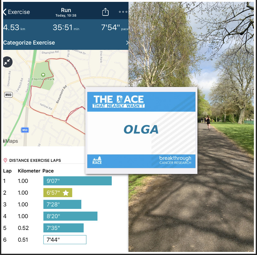
A lot has changed for me since I began my research journey in RCSI, as I transitioned from being an undergraduate placement student to a PhD candidate, however the biggest change has been adjusting to doing a lab-based PhD during a pandemic!
These days my work hours are shared between the labs in RCSI and my family home. While my bench space and office space used to be separated by just a few steps, there is now a 30+ minute bus journey between them. It has certainly put my planning skills to the test as now when I walk into the lab I need to be sure of what I am planning to do, and that I can complete the task in my pre-booked lab time slot.
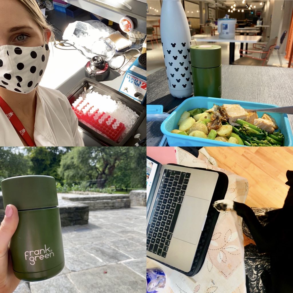
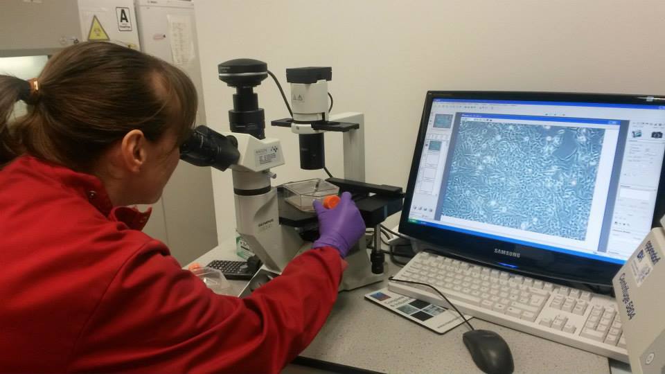
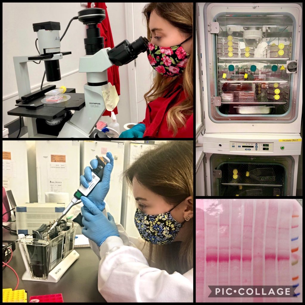
I appreciate my time in the labs much more now that I spend so much time at home. Whether I am culturing neuroblastoma cells, analyzing proteins or genes by Western blots or PCRs, I enjoy immersing myself in the work knowing that my time on the bench is limited.
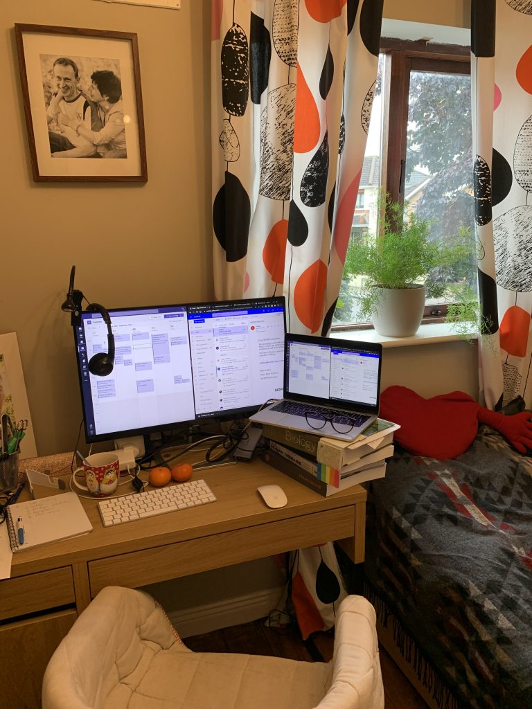
The main perk is that now when I am doing computer work – analyzing results, writing reviews, preparing presentations, using online software – I can do it from the comfort of my box-room-office, often very cosy in a blanket as I do it. While my work-from-home desk space is slightly more spacious than my desk in the now-closed “Write-up Room 2”, I do miss the chats and laughs that come with working in a shared office.
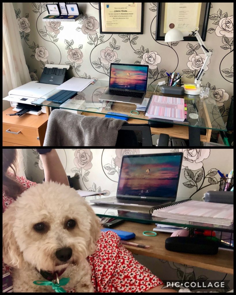
My WFH set-up. My new office buddies don’t quite understand the need for personal space.
One thing’s for sure though, my two dogs very much enjoy the days that I work from home!
Catherine Murphy, Neuroblastoma UK funded PhD student