Why do we need fundraising for cancer research?

There is no short answer. Research is a slow, meticulous process of testing theories and finding out which ones work.It is exactly the same for both curiosity- and disease- driven questions. Long years of ground research full of ups and downs are critical for any breakthrough or progress. Very often with more downs than ups. Importantly, all researchers build on the work of their predecessors. This is the nature of science.

To understand the world around us, we have to do be curious and do “blue sky or curiosity-driven” research. It is a long shot, but this type of research can lead to practical applications down the road. One of the most recent examples is a drug Vismodegib (Erivedse) to treat basal cell carcinoma (the most common type of skin cancer) approved by the FDA in 2012. This drug targets genes of a hedgehog-associated signalling pathway. Defects in this pathway were found to drive many cases of skin cancer. But, how this relationship was found? Blue sky research!

Researchers studied hedgehog signalling in fruit flies and mice. One of the researchers had a strong interest in a fruit fly gene called hedgehog. If this gene is defective, then fly embryos look stubby and hairy aka a hedgehog. Further research brought more interesting facts and relationships leading to the identification of a drug that can stop the function of this faulty gene. Decades later with the advancement of genome sequencing, the defect in hedgehog signalling pathway genes was identified in patients with locally advanced and metastatic basal cell carcinoma.

What would happen if there were no research in fruit flies and mice? There would have been no rationale to create a drug like Vismodegib!

The best discovery research is unrestricted. It is driven by intellectual curiosity and conceptual advancement. More such curiosity- driven research is needed. For every medical breakthrough, for every Vismodegib, there were hundreds of blind alleys and failed ideas.

The research is a long-term investment. This contradicts to the short-term life of the politicians and governments who give the money. They do not take the risks. So, the discovery research becomes critically underfunded.

Fundraising creates opportunities for blue sky research and developing cancer treatments.

Thank you all who support cancer research charities!


E-poster at SIOP2016

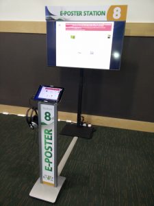
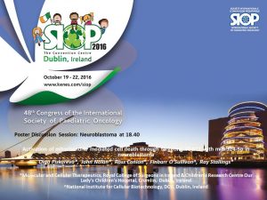
Ok. Now, when the stress of the presentation is over, I am happy to share new technologies used during the SIOP2016. As I mentioned yesterday, my work was selected for e-poster presentation. It looked this way:

This is e-poster station, where anyone can look up all posters displayed during the meeting.


It is definitely a step forward. Anyone can look up any poster, listen to a commentary recorded by the author, zoom in and out and send a request/comment to the author. It looks cool and trendy. Though, you can feel invisible as no physical copy displayed in a designated area. No crowds of poster presenters and judges. No waiting faces desperate to share their study…

The actual Poster Discussion session was a traditional presentation when my poster was up on the big screen, I had 8 minutes to convince the audience navigating through figures. This session was late and no many attendees survived to come and challenge your statements. Nevertheless, it was enjoyable experience. 🙂

siop-2016-banner-728-x-90


Last minute discovery with SIOP2016


SIOP is the International Society of Paediatric Oncology. It is a global multidisciplinary society representing doctors, nurses, other health care professionals, scientists and patients or their relatives. The Society’s motto is ‘no child should die of cancer’. The meeting 2017 is being held in Dublin, the city where I live and work.

Indeed, it was appealing to attend the key meeting in childhood oncology field. As any participant, I had an opportunity to submit an abstract about my research.  To no surprise at all, I received email notifying me on my work being selected for e-Poster presentation.  Common stuff. The email also said that it would be displayed at designated stations, like big screens throughout the meeting. Very unusual format, but we are living in the digital technology era; things are changing all the time. So, I would not need to stay by the poster this time. Great – more time for networking and talks.

Then I received another email informing about a Poster Discussion session, which I assumed to be a standard procedure when a group of selected piers stand by your poster and ask Qs. None comes in majority cases. A participant stands and waits and waits till the session is over. So, of course I took it easy.

A day before the meeting, I downloaded the meeting app and started to browse along the content and features. Out of curiosity, I checked details of the Poster Discussion session. This was the moment of mental breakdown – I discovered being selected for an oral poster presentation! My chances were 1 in 1475 (the number of submitted abstracts). I should probably also buy a lottery ticket tonight. Could lucky things come together?

I will reflect on the new e-poster presentation experience later today…


siop-2016-ppt


http://www.siop2016.kenes.com/


Drug resistant neuroblastoma cells

Children with neuroblastoma undergo several cycles of intensive chemotherapy to stop disease progression with the final aim to eliminate the tumour. Chemotherapy includes carboplatin or cisplatin in various combinations with drugs such as cyclophosphamide, ifosfamide, doxorubicin, etoposide, topotecan and vincristine (1). Nevertheless, in average 1 in 5 children with stage 4 disease do not respond to therapy. Up to 50% of children that do respond experience disease recurrence with tumour resistant to multiple drugs and more aggressive behaviour that all too frequently results in death.

The development of drug resistance is the major obstacle in treatment of neuroblastoma. To tackle this problem, researchers need to study different models of disease using cell lines, 3D tumour cell models, mice models and have access to clinical samples.

The first stage in testing drugs is to understand their killing ability of cancer cells. At this stage, researchers test drugs using cell lines. Cell lines are derived from tumours which were surgically removed from children with neuroblastoma. Researchers usually take a small piece of tumour straight after surgery and bring it into the laboratory.  Here, they place this piece into special solution that has enzymes to separate cells from each other. Then the suspension of all kind of tumour cells is placed into plastic dishes or flasks in a highly nutrient media to let cells grow. Cells that can adapt to these conditions start to grow, divide and produce a new generation of cancer cells. Researchers look after their growth, inspect their shape and behaviour; and test them on the presence of tumour markers. Once identity of these cells is confirmed they become a cell line and obtain a name. These cells keep majority of characteristics of the parental tumour and represent very useful tools in cancer research.

In our lab we use such cell lines to study neuroblastoma resistance to drugs. To understand changes in neuroblastoma biology during the development of drug resistance, we created drug resistant neuroblastoma cell lines (2). We treated three neuroblastoma cell lines CHP212, SK-N-AS and Kelly with cisplatin – a common drug in anticancer therapy. SK-N-AS and Kelly cells are sensitive to this drug, while CHP212 cells responded to this drug at much higher levels that the other two. Cells were grown in media containing cisplatin for several weeks. During this period most of the cells responded to cisplatin and died. Then we let cell survivors to recover in media without drug. This cycle was repeated several times until we got a population of cell survivors that can stand doses of cisplatin that can kill 50% of parental cells.  It took us more than 6 months to generate cisplatin resistant neuroblastoma cell lines CHP212Cis100, SK-N-ASCis24 and KellyCis83.

At the next step, we studied differences between these cell lines. We first compared their behaviour and cell shapes. Two resistant cell lines KellyCis83 and CHP212Cis100 started to grow faster, but SK-N-ASCis24 – slower than their parental cell lines. Interestingly, these cells also became more resistant to other drugs such as doxorubicin, etoposide, temozolomide, irinotecan and carmustin. These results are very important as they demonstrate that one drug can activate the cell defense systems that allow to escape toxicity of other drugs. These cell lines can be used to test new drugs and find those that can overcome developed resistance.

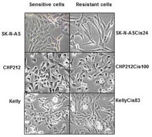
Cisplatin resistant cells also changed their appearance. Most dramatic changes occurred in SK-N-ASCis24 cells (see Figure 1).
nbl-cells

Figure 1. Microscopic images sensitive and drug resistant neuroblastoma cells (adapted from (2)) 

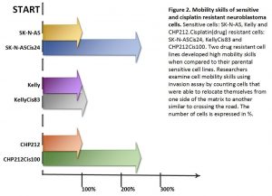
Two drug resistant cell lines SK-N-ASCis24 and CHP212Cis100 cells developed additional mobility skills – they became more invasive than their parental counterparts.
 

resistant-cells


Then we asked a question: what type of changes allowed cells to adapt to cytotoxic environment?  We examined changes in their genomic DNA first. We found that some genes increased their copy number, other went missing.

We identified changes in protein expression. More intriguingly, some proteins with the increased presence in the cells did not increase their presence in genomic DNA. We sorted these proteins on their role in cell processes such as migration, growth, cell cycle, etc. We found that each cisplatin resistant cell line developed a unique set of features that help them to escape cytotoxic stress (2). The similar patterns are found in clinic. Each patient responds to treatment differently.

What did we learn from this study?

  • One drug, in our study cisplatin, can activate the cell defense systems that allow to escape toxicity of other drugs.
  • The development of drug resistance gives cells new advantages and changes their behaviour and appearance, e.g. mobility skills, different cell shape, response to drugs, etc.
  • Each cisplatin resistant cell line developed a unique set of features that help them to escape cytotoxic stress.
  • These cell lines can be used to test new drugs and find those that can overcome developed resistance.

References

  1. Davidoff AM. Neuroblastoma. Semin Pediatr Surg. 2012; 21(1):2–14.
  2. Piskareva O, Harvey H, Nolan J, Conlon R, Alcock L, Buckley P, et al. The development of cisplatin resistance in neuroblastoma is accompanied by epithelial to mesenchymal transition in vitro. Cancer Lett. 2015;364(2):142–55. 


Cell to Cell Communicators

Tumour cells send different types of messages from one cell to another aka people post letters, postcards, and parcels to their families, friends, colleagues or  business. Cells can direct their messages using free moving proteins – postcards. They can wrap it in microvesicles with different cargo. Big microvesicles can take up big messages – parcels, small microvesicles or exosomes contain a limited number of texts – letters.

Tumour cells change their behaviour quickly adapting to anticancer therapies, so the messages they are sending. These messages can easily join blood stream and be read by researchers to understand how treatment is working and tumour cells are feeling.  Reading these messages from blood is more favourable as blood tests are done on the regular bases during and after the treatment.

In our lab we investigate how neuroblasts communicate with each other and the entire body through exosomes. We are interested to see what they write in their letters – exosomes. Do drug resistant and sensitive neuroblasts write different texts? What is the difference and how we can use this difference to predict child response to anticancer therapy?

In one set of experiments, we found that exosomes from drug resistant neuroblasts stimulate growth of sensitive cells. The more resistant neuroblasts send more powerful messages pushing cells to grow faster.

In the other set of experiments, we partially cracked the message showing that their texts are different. This finding explains why more resistant neuroblasts send more growth stimulating messages.

All these findings will be presented at the upcoming conference Goodbye Flat Biology: Models, Mechanisms and Microenvironment in Berlin.


schematic-exo2a

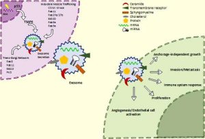
Schematic of exosome biogenesis and secretion. Cells produce exosomes through different pathways. This process is tightly regulated and controlled by numerous molecules. It can be triggered by many factors including extracellular stimuli (e.g., microbial attack, UV, drugs) and other stresses. The exosomes wrap up biologically active components such as proteins, RNA and miRNA. Exosomes can interact with recipient cells using four mechanisms: ligand/receptor interaction, protein transfer, membrane fusion or internalisation. Once exosomes entered the recipient cell, they release their content and re-programme the cell functions.


Suggested reading

Johnsen KB, Gudbergsson JM, Skov MN, Pilgaard L, Moos T, Duroux M. A comprehensive overview of exosomes as drug delivery vehicles – Endogenous nanocarriers for targeted cancer therapy. Biochim Biophys Acta – Rev Cancer. 2014;1846(1):75–87.

El Andaloussi S, Mäger I, Breakefield XO, Wood MJ a, Andaloussi S EL, Mäger I, et al. Extracellular vesicles: biology and emerging therapeutic opportunities. Nat Rev Drug Discov. 2013;12(5):347–57.

The schematic of exosomes was adapted from here