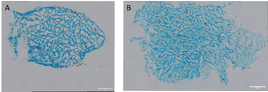
The most enthusiastic followers may remember my previous post, where we obtained some slices of our “brain-like” hydrogel to study how the cells within behave. Here we dive into the hydrogel itself by cutting it into razor-thin slices (10 μm, or one hundredth of a millimetre), so we can observe the intricate web of hyaluronic acid filaments that compose the gel and will sustain the cancer cells in our model. In the picture below, we can admire some slices obtained from 2 gels with different concentrations of Methacrylated Hyaluronic Acid (MeHA), which is the main component of the gel. The blue colour is a result of the hydrogel stained with alcian blue, which attaches the molecules of hyaluronic acid and gives this light blue.
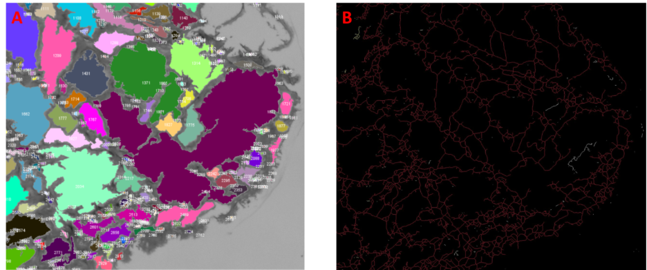
All this work is not just to get pretty pictures. This way we can measure the empty space in the gel which cells and other components can move freely, as well as how much surface the hyaluronic acid gives the cells to grow and spread. Here we can see the representation of the pores, with the measurement of the size next to the detection of the backbone (the hyaluronic acid). Smaller pores may indicate higher robustness of the gel, but lower diffusion capacity for nutrients, drugs or cells. Meanwhile, a higher number of junctions enhances network integrity and elastic recovery up to a limit, though excessively high junction densities can lead to easily breakable networks. The same technique will be used in the future to obtain sections of the hydrogel with the tumour cells growing on it to study their spreading and morphology.
Bear in mind that by cutting the gel, we are obtaining 2D images from a 3D hydrogel; thus, it is a simplification of the model itself. However, we can still learn a lot from these data about the gel properties. We could explain some of the mechanical properties observed in previous experiments, like the gel’s elasticity and stiffness, both properties that influence cell behaviour and are necessary to correctly model the brain’s specific mechanical properties.
Written by Pierluca Cancellieri, Mac4Me PhD