As many will know, the Nobel Prize for Physiology or Medicine 2023 was awarded jointly to Dr. Katalin Karikó and Dr. Drew Weissman in recognition of their contribution to the mRNA technology that laid the groundwork for the Covid-19 mRNA vaccine success. They found that replacing uridine with pseudouridine in mRNA molecules prevented over-activation of an immune response that would normally degrade and remove the mRNA. Combined with successful delivery to cells, this allowed for mRNA to be used as coding machinery to make new proteins in cells that don’t normally produce that protein. One small adjustment changed the landscape of mRNA technology and therapeutics.
Since the Nobel Prize announcement, it has come to light the many obstacles and rejections that Dr. Karikó adapted to over the course of her career and despite it all, was still awarded the most prestigious title in scientific research. Taking inspiration from this, we will run a new blog series to highlight female scientists, their work and what it is that we admire most about them. (Did you know? Only 13 of the 227 Nobel Prize Laureates for Physiology or Medicine were women!)
In her many interviews and discussions, it is obvious that Dr. Karikó has an exceptional positive mental attitude and, most importantly, lives and breathes for science. The title of this blog post, “I felt like a God”, is a quote from Dr. Karikó. Most people will think that it is a reaction to hearing she won the Nobel Prize. But no, it was Dr. Karikó’s reaction in the 1980s to seeing her mRNA inserted into cells and producing a new protein. Dr. Karikó was still working at the bench up until very recently, and this love for lab work and science is one quality that makes her stand out from the crowd. She shy’s away from the “fame” and focuses on “what’s next”. It is these qualities, I assume that meant she could push past the rejections that we know so well in academia and scientific research. Before her and Dr. Weissman’s work was noticed and valued by Pfizer and Bio-N-Tech, Dr. Karikó was let go from the University of Pennsylvania due to a lack of grant funding and struggled to attract interest from both industry and academia. In fact, major journals rejected Dr. Karikó and Dr. Weissman’s work on mRNA modifications, and it was finally published in a lesser-known journal, Immunity.
Rejections are something that every scientist becomes accustomed to, be they grant rejections, job rejections or even “idea” rejections. Dr. Karikó’s ability to firmly believe in her science and never let up is what I admire most about her. She was told so many times to “not bother” with RNA that it would never be used as a therapeutic. And now, her belief and self-confessed obsession with this molecule has contributed to the development of arguably the most ground-breaking therapeutic of the 21st century, the mRNA vaccine. Obviously, as an immunologist, I am biased. But it cannot be ignored how many lives Dr. Karikó’s ambition and determination have benefited.
As women in science, we face unique challenges. Dr. Karikó’s career is not what you would expect from a Nobel prize-winning scientist. It is not littered with tenured positions at Ivy League universities. She struggled for most of her career to get people to listen to her about the promise of mRNA. Moving from her home of Hungary to the United States with her husband and young daughter, Susan, who is now a two-time Olympic gold medallist. When asked how she juggled it all, Dr. Karikó explains how leading by example is the key. It is no wonder her daughter is as successful as she is when Dr. Karikó’s determination, perseverance and self-confidence were what she grew up watching. I truly admire Dr. Karikó’s ability to stand firm in the face of adversity. For me, she stands as a true role model for women scientists and embodies the success that resilience can bring.
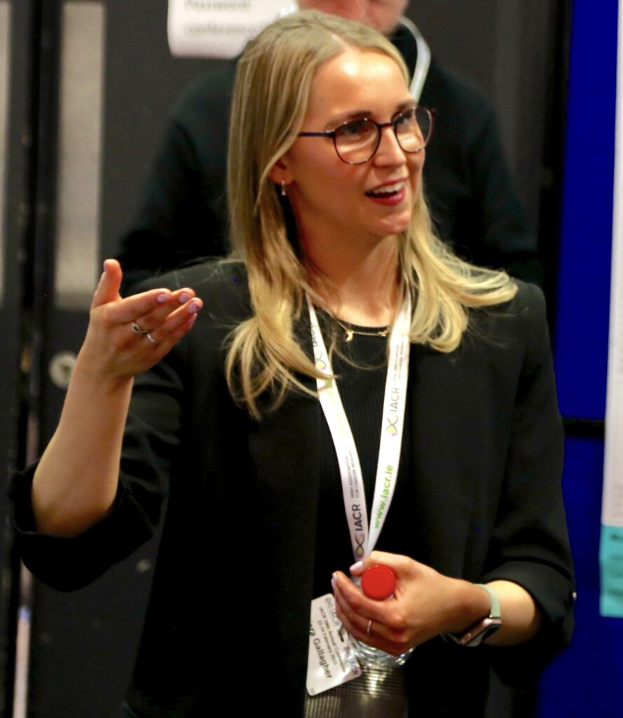
Written by Ellen King